Where is the best place to teach joint ultrasound studies? FGBU FNKTS FMB of Russia
In practice, ultrasound of joints began to be used not so long ago, but this has expanded the possibilities for diagnosing diseases of large and small joints. The test is called this because the machine works by passing ultrasound waves at a specific frequency through tissue. Ultrasound techniques are used in many branches of medicine, for example, traumatology or orthopedics, which facilitates the diagnostic process not only in intra-articular structures, but also in tissues and nerves.
Ultrasound of joints can be performed if destructive processes in tissues are suspected.
Advantages and disadvantages
Ultrasound of joints is a non-invasive imaging method with which the doctor examines the condition of the skeleton. This method is not as expensive as others and allows you to get results quickly. The disadvantages of this procedure include the need for special training of medical staff and the need to purchase special high-quality equipment. Otherwise, ultrasound examination of joints is a safe and informative way to diagnose many diseases. Ultrasound shows detailed information on a wide range of diseases.
When is it held?
The study of bones allows us to record all the processes that occur on the surfaces of bone tissue. Ultrasound of bones and joints is prescribed when:
- back pain;
- suspicion of dysfunction of large joints, including knees, shoulders, elbows;
- limb injuries;
- arthritis and arthrosis;
- cyst and tumors;
- dislocations and subluxations of the hip;
- dysplasia.
Preparation for the event
 An ultrasound scan of joints does not require special preparation.
 The procedure itself is carried out in a separate room without prior preparation on the part of the patient. The examination is painless and can be done in 15 minutes. Ultrasound does not affect the general condition of a person in any way. The result can be obtained instantly, the diagnostician issues a conclusion immediately after completion.
Absolute contraindications to ultrasound of joints are considered to be situations where waves can worsen the prognosis of the disease. The specialist lacks special education. In case of damage to the skin, it is also better to postpone the examination.
General picture of ultrasound diagnostics
How is the study of the elbow and shoulder joint performed?
Standard ultrasound of the elbow joint is prescribed for athletes who are more susceptible to sports injuries or illnesses. The procedure shows the condition of the ulnar capsule, the tendon of the brachialis and forearm muscles, cartilage, and gives an idea of the condition of the radial and ulnar nerves. In turn, the shoulder joint is studied while sitting on a chair. During the procedure, the doctor will change the position of the required area for better visibility on the screen. Using a shoulder examination, it is possible to examine the subscapularis and infraspinatus muscles, the joint capsule, tendons and intra-articular cartilage.
Ultrasound of the mandibular joint and wrist joint
When diagnosing pathologies of the temporomandibular joint (TMJ), ultrasound is preferable to MRI. The study of the TMJ allows us to examine the meniscus, capsular-ligamentous and muscular apparatus. The algorithm for studying the jaw joint is based on examination first with the mouth open and then closed. The next type of study makes it possible to diagnose pathology of the carpal tendon, blood vessels, muscles and nerves. In addition, the soft tissue of the finger or fingers is studied. Ultrasound of the wrist joint detects fractures of the hands, which cannot always be detected by x-rays, and the effectiveness of the study is no worse than the MRI technique.
Knee ultrasound is a type of ultrasound examination that allows visualization of the knee joint. This study helps differentiate between diseases such as rheumatoid arthritis, traumatic injuries and degenerative pathologies.
Often the pathological process can be detected by ultrasound at an early stage, due to which treatment in many cases is successful. This test is safe and can be performed even on a child.
Where to do the study - this study is carried out by all ultrasound doctors and even orthopedists-traumatologists in ultrasound diagnostic rooms.
How to prepare for the procedure
No preliminary preparation for the procedure (hunger, drinking water, etc.) is required. But the study should not be performed within four to five days after intra-articular injections.
Research methodology
Scanning of the anterior and lateral parts of the knee joint is carried out with the patient lying on his back, with his knees extended. To better visualize the intra-articular cartilage (menisci), the patient is asked to bend his knees.
To examine the posterior parts of the joint, the patient is turned onto his stomach. After the examination, the patient receives a research protocol with a conclusion.
Sonography is carried out in several projections, here are some of them:
1.Anterior longitudinal projection
The sensor is placed above the patella parallel to the femoral axis. In this position, the hyperechoic patella with the femur is examined, which is also visualized as a linear hyperechoic structure.
Between them the suprapatellar bursa is visible in the form of a triangle, located under the tendon of the quadriceps femoris. When the sensor is moved below the patella, the patellar ligament, tibia, infrapatellar bursa and fat pad are visualized.
2.Anterior transverse projection
The sensor is placed over the upper edge of the patella. In this case, the femur and hyaline cartilage are visualized (a hypoechoic uniform strip), the suprapatellar bursa is visible in cross-section. The synovium in a healthy person is not visualized. When the sensor is moved downward, the anterior condyles of the tibia are visible.
Read also:
7 indications for ultrasound examination of the mammary glands
3.Posterior transverse projection
The examination takes place lying on your stomach. Thus, the posterior condyles of the femur, hyaline cartilage and up to the popliteal fossa are visualized.
4.Posterior lateral projection
The condyles of the femur and tibia, the intercondylar joint space, and the posterior horn of the lateral meniscus are examined.
5. Posterior longitudinal medial projection
The medial part of the popliteal fossa with the condyles of the femur and tibia, the joint space, cartilage, the posterior horn of the medial meniscus, and the semimembranosus tendon are examined.
That a doctor’s visit is normal and in case of pathology
Norm
Normally, an ultrasound of the knee joints examines all structures. In this case, the articular surfaces should be smooth, clear, without deformations, there should be no effusion in the articular cavity, the synovial membrane should not be visible, and the articular capsules and inversions will look like hypoechoic formations of a folded structure with multiple branches. Hyaline cartilage should be homogeneous in structure.
Pathology
Patients are often interested in what an ultrasound scan of the leg joints shows. After all, this is not the stomach, which we are already accustomed to scanning. So, this type of research helps to determine the disease with 99.9% accuracy. These include ligament ruptures, arthritis, complex intra-articular fractures, and other pathologies.
All pathological changes in the knee joint can be divided into groups:1. Traumatic damage to the tendon-ligamentous apparatus of the joint (rupture of the patellar ligament or quadriceps tendon, microtraumatization of the lateral ligament, etc.).
Injuries to the ligamentous apparatus occur due to a fall, inadequate load on the joint and awkward sudden movements. In this case, complete ruptures look like a complete violation of the integrity of the tendon, with the appearance of hypoechoic areas between the ends of the damaged ligament. Hematomas may be detected. 
With a partial rupture, the contours of the tendon are preserved, but a hypoechoic area is clearly visible at the site of damage. When the patella is fractured, a hypoechoic gap is visualized between the bone fragments. In case of polytrauma, examination of the ankle joint is indicated.
Read also:
We determine the condition of the leg veins using duplex scanning
2. Pathology of the menisci, condition after surgery. The main signs of meniscal damage are disruption of the contour line of the damaged meniscus, the formation of hypoechoic areas and stripes, effusion in the articular cavity, signs of edema, and displacement of the collateral ligaments. Degenerative changes in the menisci look like structural heterogeneity.
3. Degenerative process, for example, Pellegrini-Stied calcification, in which multiple ossifications are identified as hyperechoic formations in the ligamentous apparatus of the knee joint. With deforming arthrosis, a narrowing of the joint space is detected, the hyaline cartilage is unevenly thinned, the contours of the articular surfaces are uneven with osteophytes.
4.Dysplastic process.
5.Cysts are visualized as delimited cavities filled with fluid.
6. Inflammation of the synovial membrane, its hyperplasia, osteochondromatosis, villonodular synovitis, synovial sarcoma, as well as rheumatic synovitis. An inflammatory process is indicated by effusion in the joint cavity.
7.Tendinitis, in which the echo density of the tendons decreases.
Features of the study, price

The main feature of the study is its accuracy and reliability, but the doctor performing the study must have a good knowledge of the anatomy of the knee joint. Often, in case of combined injuries, an ultrasound of the ankle joint is also performed.
The scan can be done in a city clinic equipped with special equipment, or in any specialized diagnostic center. This test can be done for a child at a children's orthopedic clinic. The cost of the procedure varies from 600 to 2000 rubles.
Educational process
The excellent material base of the department - the diagnostic departments of the Federal Scientific and Clinical Center FMBA - are dozens of complex modern expert-class diagnostic devices: modern ultrasound scanners, high-field magnetic resonance imaging scanners from the world's leading manufacturers (Siemens, General Electric, Philips), multi-slice computed tomographs, stationary and mobile digital x-ray equipment . Technical equipment makes it possible to train doctors at the modern level in various areas of radiation diagnostics in cycles of improvement in the following topics: CT and MRI diagnostics of various organs and systems (diagnosis in rheumatology, diagnosis of diseases of large and small joints, differential diagnosis of diseases of the lungs, liver and biliary tract ), ultrasound examinations of the heart, including modern diagnostic methods (Doppler echocardiography, stress echocardiography, transesophageal echocardiography), ultrasound examination of blood vessels, ultrasound diagnostics of the musculoskeletal system.
Particular attention is paid to the cycle of professional retraining in ultrasound diagnostics. Training is provided in methods of studying organs and organ systems of the abdominal cavity, pelvis, retroperitoneal space, blood vessels, heart, joints, thyroid gland and superficial organs in accordance with the approved program. The form of presentation of the material is lectures, practical classes, seminars, master classes with the participation of experienced teachers and practitioners.
The cycle of advanced training for radiologists (certification) includes current issues of classical x-ray diagnostics, at the same time, the emphasis is on highlighting the capabilities of modern methods of radiological diagnostics: multi-slice computed tomography, magnetic resonance imaging, digital mammography, x-ray osteodensitometry. Cadets are introduced to the latest areas of radiation diagnostics: MRI of joints, MRI of the heart, MSCT of large vessels, complex radiation diagnostics of diseases of the central nervous system.
The department trains radiographers in the special training cycle “Laboratory Science in Radiology”, graduating practitioners trained in practical skills of working with the most modern technology (CT, MRI, mammography, densitometry, orthopantomography).
A separate area of educational activity of the department is the training of high-level specialists through clinical residency in radiology and ultrasound diagnostics. Experienced teachers work in this area - recognized practitioners known in the country and abroad. The high level of training of clinical residents is the key to the demand for young specialists in the labor market. Every year, the number of people wishing to study for residency at the Department of Radiology and Radiation Diagnostics exceeds the capacity of the clinical base.
The department carries out extensive scientific work in various areas of radiation diagnostics. The department's annual scientific output includes dozens of publications in the country's leading scientific journals and in foreign publications. During the work of the department, more than 2000 cadets from many cities of Russia and CIS countries (Ukraine, Belarus, Kazakhstan, Moldova, Armenia, etc.) were trained.
Retraining in the specialty “Ultrasound Diagnostics” (USD), general improvement in ultrasound diagnostics (certification cycle).
In recent years, there has been extraordinary progress in computer technologies and their accelerated introduction into everyday medical practice. At the forefront of medical imaging is ultrasound diagnostics of diseases of various organs and systems of the human body. Ultrasound, due to its high information content, non-invasiveness, speed of execution, and the possibility of repeated repetition without harm to the patient’s health, is recognized not only as a search method, but also as a method of choice in the diagnosis of many diseases. Technological innovations in recent years have allowed ultrasound diagnostics to reach a completely new qualitative level, which in modern conditions has required new approaches and knowledge from medical specialists.
At the Department of Radiology and Ultrasound Diagnostics of the Academy of Postgraduate Education of the Federal State Budgetary Institution Federal Scientific and Clinical Center of the Federal Medical and Biological Agency of Russia, doctors are trained in a cycle of professional retraining in ultrasound diagnostics in accordance with the program and educational standards. The form of presentation of the material is lectures, practical classes, seminars, master classes with the participation of experienced teachers and practitioners. The presentation of the topics studied and the conduct of practical training takes into account the different levels of initial knowledge of doctors. Cadets are taught the clinical basis of diseases of internal organs, the theory and methodology of ultrasound examinations, study methods for studying organs and systems of the abdominal cavity, pelvis, retroperitoneum, blood vessels, heart, joints, thyroid gland and superficial organs. Along with theoretical knowledge, an important aspect is obtaining practical skills in conducting ultrasound techniques during practical classes in the offices of the Department of Functional and Ultrasound Diagnostics of the Federal Scientific and Clinical Center of the Federal Medical and Biological Agency of Russia under the guidance of experienced teachers. The topics of the cycle introduce cadets to both the classical ultrasound semiotics of the main nosological forms and the new capabilities of ultrasound in the study of diseases of the musculoskeletal system and lymph nodes; the latest technological techniques are presented - ultrasound angiography and power Doppler. The duration of the primary training cycle (PP) “Ultrasound diagnostics” for doctors of medical specialties who graduated from a university before 2000 is 4 months (total number of training hours - 576). The duration of the general improvement (GP) cycle “Ultrasound diagnostics” for ultrasound diagnostic doctors is 4 weeks (total number of training hours - 144). Lectures and practical classes on the courses are conducted by professors, doctors of medical sciences Marina Albertovna Chekalova, Ekaterina Mikhailovna Nosenko, candidates of medical sciences Olga Vladimirovna Pushkova, Natalya Sergeevna Nosenko, Tatyana Vladimirovna Grandfathers. At the end of the cycle, control tests are offered and an exam is held in the form of an interview, based on the results of which cadets of the PP ultrasound diagnostics cycle receive a diploma and certificate of an ultrasound diagnostics specialist, cadets of the OU ultrasound diagnostics center receive a certificate of completion of the cycle and a certificate of an ultrasound diagnostics specialist.
General improvement in the specialty “Radiology” (certification cycle).
At the Department of Radiology and Ultrasound Diagnostics of the Academy of Postgraduate Education of the Federal State Budgetary Institution Federal Scientific and Clinical Center of the Federal Medical and Biological Agency of Russia, training of radiologists is carried out on the cycle of general improvement “Radiology” for radiologists who have primary education in the specialty and a specialist certificate. The training lasts 6 weeks (total number of training hours - 216). Classes are held on the basis of the X-ray department with magnetic resonance imaging rooms of the Federal State Budgetary Institution Federal Scientific and Clinical Center of the Federal Medical and Biological Agency of Russia. Training is conducted at the level of advanced global and domestic requirements. The presentation of the topics studied and the conduct of practical classes takes into account the different levels of background knowledge of cadets. The advanced training cycle includes current issues of classical x-ray diagnostics, while the emphasis is on highlighting the capabilities of modern methods of radiological diagnostics: multi-slice computed tomography, magnetic resonance imaging, digital mammography, x-ray osteodensitometry. Cadets are introduced to the latest areas of radiation diagnostics: MRI of joints, MRI of the heart, MSCT of large vessels, MSCT of the heart and coronary arteries, and issues of complex radiation diagnosis of diseases of the central nervous system are studied. Expert-class equipment is used in the educational process - multifunctional digital X-ray complexes, superconducting magnetic resonance imaging scanners, multispiral computed tomographs, dual-energy osteodensitometer and much more. Lectures and practical classes on the cycles are conducted by: Head. Department of Radiology and Ultrasound V. N. Lesnyak, professors, doctors of medical sciences A. V. Smirnov, E. B. Guzeeva, candidates of medical sciences E. O. Kontarova, E. A. Zvezkina. An important direction is to gain skills in carrying out the studied techniques in practical classes with doctors from the radiology department with computer and magnetic resonance imaging rooms. Upon completion of training, cadets receive a standard certificate and a specialist certificate.
Ultrasound diagnosis of heart diseases (standard basic echocardiography) - thematic improvement (TU)
At the Department of Radiology and Ultrasound Diagnostics of the Academy of Postgraduate Education of the Federal State Budgetary Institution Federal Scientific and Clinical Center of the Federal Medical and Biological Agency of Russia, training of ultrasound diagnostic doctors, functional diagnostic doctors, and medical doctors in the technique of transthoracic echocardiography is carried out. The training lasts 3 weeks (108 hours) and includes a theoretical part - lectures that provide information about the structure and function of the heart under normal conditions and in various pathologies. Particular attention is paid to modern global and domestic requirements for assessing heart function, taking into account the development of cardiology and cardiac surgery. The practical part includes master classes where cadets become familiar with the technique of conducting echocardiography, as well as practical exercises. Practical classes provide an opportunity for all students to master the technique of transthoracic echocardiography under the guidance of an experienced specialist, and to evaluate the features of conducting research on devices of different levels. Classes are conducted at the Center for Ultrasound and Functional Research Methods of the Federal State Budgetary Institution Federal Scientific and Clinical Center of the Federal Medical and Biological Agency of Russia by experienced teachers and highly qualified specialists with extensive practical experience: associate professors T.V. Krutova, S.N. Safronov. At the end of the cycle, a state certificate is issued.
Cycles of thematic improvement (CME) “Transesophageal echocardiography in a hospital setting and in cardiac surgery practice”, “Stress echocardiography”.
The cycles introduce students to the most modern methods of ultrasound diagnostics of heart diseases, allowing them to acquire not only theoretical knowledge, but also practical skills in these areas. Classes are conducted on the basis of the Department of Functional and Ultrasound Diagnostics of the Federal State Budgetary Institution Federal Scientific Center of Federal Medical and Biological Agency by experienced teachers: associate professors S.N. Safronov, T.V. Krutova. The cycle programs include lectures on the main topics of ultrasound examination of the heart in normal conditions and in various cardiovascular pathologies. Forms of training include a variety of illustrative material (computer display, visual teaching aids, videos, etc.). Lectures and seminars cover issues of ultrasound diagnostics of heart diseases in comparison with the clinic and the results of other instrumental research methods, and also discuss issues of indications for surgical and endovascular treatment, ultrasound monitoring of these methods of exposure. Training is conducted using modern expert-class ultrasonic equipment from leading foreign companies. At the end of the cycle, control tests are offered and an examination is conducted in the form of an interview. The duration of the internship “Ultrasound diagnosis of heart diseases (transthoracic, standard basic echocardiography)” is 3 weeks (total number of training hours - 108). The duration of the thematic improvement cycle “Transesophageal echocardiography in a hospital setting and in cardiac surgery practice” is 36 training hours, the cycle “Stress echocardiography” is 18 hours. At the end of the cycles, documents of the established form are issued.
Ultrasound examination of blood vessels (color duplex scanning of the arterial and venous systems) - thematic improvement (TU).
At the Department of Radiology and Ultrasound Diagnostics of the Academy of Postgraduate Education of the Federal State Budgetary Institution Federal Scientific Center FMBA of Russia, the author's course of Doctor of Medical Sciences, Professor Ekaterina Mikhailovna Nosenko is being conducted. The training is conducted at the Center for Ultrasound and Functional Research Methods of the Federal State Budgetary Institution Federal Scientific and Clinical Center of the Federal Medical and Biological Agency of Russia for 3 weeks (108 hours). The cycle is intended for doctors of ultrasound and functional diagnostics, medical specialties and includes lectures, master classes and practical classes with the development of practical skills, illustrative materials are provided. The course includes information on the full spectrum of vascular pathology of the head (including transcranial and ocular vascular studies), extremities, abdomen, retroperitoneum and pelvis. Aspects of therapeutic and surgical pathology, features of studies of the vascular bed are covered, taking into account modern requirements. The cycle provides an opportunity for all students to master the methods of studying the vascular system used in real practice. The presentation of the topics studied and the conduct of practical classes takes into account the different levels of initial knowledge of cadet doctors. The most important thing is to gain practical skills in conducting color duplex scanning techniques with color flow mapping at the professor’s master classes and practical classes by doctors of the highest category of the department. At the end of the cycle, a state certificate is issued.
Ultrasound diagnosis of joint diseases - CME cycles “Ultrasound diagnosis of joints of the upper extremities”, “Ultrasound diagnosis of joints of the lower extremities”.
In the arsenal of modern doctors there is a whole range of instrumental radiation methods for studying the musculoskeletal system. Together with traditional X-ray examination of bones and joints, highly informative diagnostic methods such as ultrasound (US), computed tomography (CT) and magnetic resonance imaging (MRI) are also widely used. At the same time, modern ultrasound is becoming the most suitable method for the fastest and most accessible radiological diagnosis of changes in both soft tissues and joints. Thanks to new ultrasound devices using all the achievements of modern computer technology, it has become possible to conduct a highly informative study of the musculoskeletal system for various injuries and diseases of muscles, ligaments, tendons and cartilage tissue. Doppler techniques (color and energy mapping) provide assessment of the vascular response in the area of detected changes and are used for ultrasound monitoring of treatment results. All this explains the increased interest of specialists in ultrasound examination of the musculoskeletal system. Cadets are taught the clinical basis of diseases of the musculoskeletal system, the theory and methodology of ultrasound examinations. Forms of training include a variety of illustrative material (computer display, visual teaching aids, videos, etc.).
The main lecturer of the course “Ultrasound diagnosis of joint diseases” is Candidate of Medical Sciences, Associate Professor Olga Vladimirovna Pushkova. The duration of each cycle is 36 training hours. After completing the cycle, cadets are issued a certificate of completion of thematic improvement of the state standard.
“Laboratory work in radiology” - special training, “Laboratory work in radiology” - general improvement.
At the Department of Radiology and Ultrasound Diagnostics of the Academy of Postgraduate Education of the Federal State Budgetary Institution Federal Scientific Center FMBA of Russia, radiology technicians, graduates of medical schools and nurses are trained in laboratory work in radiology, in the specialization cycle - “Laboratory work in radiology” lasting 3 months (432 hours). The cycle of general improvement (GP) “Laboratory work in radiology” for x-ray technicians lasts 6 weeks (total number of training hours - 216).
During the training, cadets of the retraining cycle become familiar with the physical foundations of the basic methods of radiological diagnostics, the problems of radiation safety of patients and personnel, and gain the necessary knowledge in the field of X-ray technology. Particular attention is paid to medical professional aspects of education: normal x-ray anatomy, basic anatomy in CT and MR images, concepts of x-ray semiotics of diseases of various organs and systems. Cadets receive practical skills in positioning for radiographic examinations, learn how to perform contrast fluoroscopic procedures and special studies. Separate areas of educational activity include training in work in mammography, x-ray osteodensitometry, computed tomography, and magnetic resonance imaging rooms. A special section of the cycle is obtaining hard copies of medical diagnostic images (classical photo process, working with modern medical printers). Classes are conducted by leading teachers of the department - head. department V.N. Lesnyak, associate professor E.O. Kontarova, assistant M.N. Kochanova, practical skills are acquired with the participation and under the supervision of the most experienced doctors and x-ray technicians of the X-ray department of the Federal State Budgetary Institution Federal Scientific and Clinical Center of the Federal Medical and Biological Agency. The cycle ends with passing internal final exams. A diploma and specialist certificate are issued.
Students of the OU cycle, along with the issues provided for in the training program, receive additional knowledge in the field of the latest digital technologies in radiation diagnostics, the basics of storing, processing and transmitting medical images. After completing the cycle, cadets receive a certificate and a specialist certificate.
Cycles on the topic of radiological diagnosis of respiratory diseases: “CT of the lungs: rare and diagnostically difficult diseases”, “CT in the diagnosis of idiopathic interstitial pneumonia” (IIP). “CT in the diagnosis of lung lesions in systemic connective tissue diseases” (CME).
Diagnosis of diffuse parenchymal lung diseases is one of the most difficult sections of radiation diagnostics. Cycles dedicated to MSCT diagnosis of lung diseases introduce students in detail to the etiology, pathogenesis, epidemiology, and clinical picture of many diseases accompanied by diffuse interstitial lung lesions. Among them are idiopathic interstitial pneumonia, lung lesions in rheumatic diseases, histiocytosis, lymphangioleiomyomatosis, bronchioloalveolar cancer, chronic eosinophilic pneumonia, exogenous allergic alveolitis, alveolar hemorrhagic syndrome, pulmonary alveolar proteinosis, pulmonary amyloidosis and others. The emphasis in presenting the material is on diagnostic features with an emphasis on the symptoms of computed tomography, the subtleties of differential diagnosis. The audience includes radiologists, as well as doctors of other specialties (pulmonologists, therapists, thoracic surgeons). The cycles are the original courses of the head of the department, V.N. Lesnyak. Duration of training on the cycle: “CT of the lungs: rare and diagnostically difficult diseases” - 72 hours, on the CME cycles “CT in the diagnosis of idiopathic interstitial pneumonia”, “CT in the diagnosis of lung lesions in systemic connective tissue diseases” - 36 hours. After Upon completion of training, cadets are issued a certificate of completion of thematic improvement of the state standard.
CT and MRI diagnostics of large joints: cycles “CT and MRI diagnostics of diseases of the joints of the upper extremities” (CME), “CT and MRI diagnostics of diseases of the joints of the lower extremities” (CME)
The computed x-ray tomography method has long and reliably established itself as the most accurate technology for assessing the condition of bone tissue. In recent years, magnetic resonance imaging has come to the fore in the study of the structures of large and small joints, due to the possibility of detailed imaging of muscles, tendons, ligaments, the condition of the synovium and bone structures. The obtained CT and MRI data form the basis of the diagnostic conclusion, which allows clinicians to determine patient management tactics, including planning surgical treatment. The topics of the cycles include current issues of traumatic injuries of large joints, diagnosis of degenerative, inflammatory diseases, tumor lesions, stress reactions. Practical classes are conducted in 3 MRI rooms equipped with high-field MRI scanners with magnetic field strengths of 1.5 T and 3 T. The material is presented in the form of lectures, analysis of clinical cases at workstations, direct work of cadets in MRI rooms under the guidance of a teacher. The contingent of students are radiologists, as well as doctors of other specialties (traumatologists, orthopedists, rheumatologists). Each training cycle has a duration of 36 hours, the trainees are issued state-issued thematic improvement certificates.
| Full name | Academic degree | Academic title | Job title |
| ZABOLOTSKAYA Natalia Vladlenovna | Doctor of Medical Sciences | associate professor | Professor |
| LELYUK Svetlana Eduardovna | Doctor of Medical Sciences | professor | Professor |
| RYBAKOVA Marina Konstantinovna | Doctor of Medical Sciences | Professor | |
| BATAEVA Roza Saidovna | Candidate of Medical Sciences | Associate Professor | |
| BRYUKHOVETSKY Yuri Anatolievich | Candidate of Medical Sciences | associate professor | Associate Professor |
| NAUMOVICH Elena Grigorievna | Candidate of Medical Sciences | Associate Professor | |
| FEDOROVA Evgenia Viktorovna | Candidate of Medical Sciences | associate professor | Associate Professor |
| SALTYKOVA Victoria Gennadievna | Doctor of Medical Sciences | professor |
The Department of Ultrasound Diagnostics of the Russian Medical Academy of Postgraduate Education of the Ministry of Health of the Russian Federation was founded on February 1, 1992 and was the first specialized department in the post-Soviet space. The Department of Ultrasound Diagnostics was separated from the Department of Medical Radiology (the head of the department is Professor Yu.N. Kasatkin).
And this is how the department was born. In 1987, the USSR Ministry of Health approved the curriculum for the training of ultrasound diagnostic specialists, developed at the Department of Medical Radiology. And then it begins a cyclic training of doctors in ultrasound diagnostics with the involvement of the All-Russian Scientific Research Center of the USSR Academy of Medical Sciences (S.A. Balter), 4th State Institution of the USSR Ministry of Health (A.V. Zubarev) and the Research Institute of Coloproctology of the Ministry of Health of the Russian Federation (L.P. Orlova). The coordinating function is assigned to TsOLIUv.
In 1989, N.V. came to the position of assistant at the Department of Medical Radiology. Zabolotskaya, and in 1991 - A.N. Khitrova. In 1990, Yu.A. entered clinical residency. Bryukhovetsky, since 1991 he has been taking part in training. In 1992 Yu.A. Bryukhovetsky becomes an assistant at the department.
At the time of its creation (01.02.92), the department included 5 full-time teaching staff (head of the department - 1, assistants - 4) and 1 teaching and support staff (senior laboratory assistant). By this time, the department carried out both cycles of professional retraining and thematic improvement. In 1992, M.V. came to the department. Medvedev, A.I. Sokolov, M.N. Skvortsova and E.V. Fedorov, in 1993 - E.G. Naumovich. Over the years, M.A. also worked at the department. Osipov (1994-2000), M.D. Musaeva (Mitkova) (1996–1999), G.G. Rudko (1996–1998), B.I. Zykin (1996–1998), O.V. Proskuryakova (1997–1999), I.A. Ozerskaya (2003-2009).
In 1992-1996. 104 training cycles were conducted. 2913 students were trained. In 1997-2001 124 training cycles were conducted. 3,459 students were trained. In 2002-2006 97 training cycles were conducted. 4071 students were trained. In 2007-2011 98 training cycles were conducted. 4109 students were trained. In 2012-2013 39 training cycles were conducted. 2308 students were trained.
In total, from 1992 to 2013, the department conducted 462 training cycles and trained 16,860 (14,552 until 2012) students.
For the period 2009-2013. Employees of the Department of Ultrasound Diagnostics published 48 articles and 37 abstracts of reports in leading specialized peer-reviewed journals, presented 105 reports at Russian and international symposia, conferences and congresses.
The department defended 11 dissertations for the degree of doctor and 40 candidates of medical sciences. 6 more works are being carried out for the academic degree of Doctor and Candidate of Medical Sciences.
All employees of the department are editors or members of the editorial board of the journal “Ultrasound and Functional Diagnostics”, and also participate in the work of other specialized journals.
Zabolotskaya N.V. and Rybakova M.K. are members of the Executive Committee of the Russian Association of Ultrasound Diagnostics in Medicine. Mitkov V.V. is the President of the Association, and Saltykova V.G. - executive secretary.
The department has developed and approved by the Ministry of Health of the Russian Federation: a unified and standard training program for ultrasound diagnostic doctors; 14 manuals for doctors; qualification tests in ultrasound diagnostics (2, 3 and 4 generations); clinical residency training program in ultrasound diagnostics.
In 1996-1998 The five-volume “Clinical Guide to Ultrasound Diagnostics” (volumes 1-5) (Vidar) was completed, in the creation of which not only members of the department, but also many leading specialists from Russia and the CIS countries participated. Later, the first three volumes also appear on CDs. In 2003, “A Practical Guide to Ultrasound Diagnostics” appeared. General ultrasound diagnostics” (edited by V.V. Mitkov) (M., Vidar, 2003). These books were the main teaching aids for several generations of ultrasound doctors and are still in demand today.
In 1997-2009 Department employees publish a number of monographs:
- “Differential ultrasound diagnostics in obstetrics” (M.V. Medvedev, E.V. Yudina), M., Vidar, 1997
- “Ultrasound mammography” (N.V. Zabolotskaya, V.S. Zabolotsky), M., Storm, 1997
- “Differential ultrasound diagnostics in gynecology” (M.V. Medvedev, B.I. Zykin, V.L. Khokholin, N.Yu. Struchkova), M., Vidar, 1997
- “Ultrasound angiology” (V.G. Lelyuk, S.E. Lelyuk), M., Realnoe Vremya, 1999
- "Dopplerography" ed. V.V. Mitkova (M.I. Ageeva, Yu.A. Bryukhovetsky, N.V. Zabolotskaya, V.V. Mitkov, M.D. Mitkova, Yu.N. Chereshneva), M., Vidar, 1999
- “Dopplerography in the diagnosis of diseases of the liver, gall bladder and pancreas” (V.V. Mitkov), M., Vidar, 2000
- “Doppler study in obstetric practice” (M.I. Ageeva), M., Vidar, 2001
- “Terminological Dictionary” (V.V. Mitkov, Yu.A. Bryukhovetsky, N.V. Zabolotskaya, E.A. Zubareva, M.K. Rybakova), M., Medica, 2003
- “Ultrasound angiology” (V.G. Lelyuk, S.E. Lelyuk), M., Realnoe Vremya, 2003
- “Ultrasound research methods in neonatology (ed. L.I. Ilyenko, E.A. Zubareva, V.V. Mitkov) M., Expressprint, 2003
- “Neurosonography in young children” Minsk, Paradox, 2004
- “Cerebral circulation and blood pressure” M., Realnoe Vremya, 2004
- “New technologies of ultrasound mammology” (N.V. Zabolotskaya, V.S. Zabolotsky), M., STROM, 2005
- “Echography in gynecology” (I.A. Ozerskaya), M., Medica, 2005
- “Reproductive system of a girl, teenager, girl” (I.A. Ozerskaya, N.V. Zabolotskaya, M., Vidar, 2007
- “A practical guide to ultrasound diagnostics. Echocardiography” (M.K. Rybakova, M.N. Alekhin, V.V. Mitkov), M., Vidar, 2008
- “Chronic pelvic pain in women of reproductive age” (I.A. Ozerskaya, M.I. Ageeva), M., Vidar, 2009
- “New technologies of ultrasound mammography.” (practical guide), N.V. Zabolotskaya, V.S. Zabolotsky, Ed. "Strom" Moscow 2010.
- “Echocardiography in tables and diagrams. Desk reference book." M.K. Rybakova, V.V. Mitkov. Ed. 1st. M.: Vidar, 2010.
- “Echocardiography in tables and diagrams. Desk reference book." Rybakova M.K., Mitkov V.V. Ed. 2nd. M.: Vidar, 2011.
- “Ultrasound diagnosis of diseases of the veins of the lower extremities” (A.R. Zubarev, V.Yu. Bogachev, V.V. Mitkov), M., Vidar, 1999.
- “Ultrasound diagnosis of diseases of the external genital organs in men” (A.R. Zubarev, M.D. Mitkova, M.V. Koryakin, V.V. Mitkov), M., Vidar, 1999.
- “Dopplerography of perinatal brain lesions” (E.A. Zubareva, I.V. Dvoryakovsky, A.R. Zubarev, A.B. Sugak), M., Vidar, 1999.
- "Dopplerography in gynecology" ed. B.I. Zykina, M.I. Medvedeva (S.E. Lelyuk), M., Realnoe Vremya, 2000
- "Guide to Diagnostic Imaging" ed. Sh.Sh. Shotemore (M.K. Rybakova), M., Soviet sport, 2001
- “Cerebrovascular reserve in atherosclerotic lesions of the brachiocephalic arteries” (S.E. Lelyuk), Kyiv, Ukrmed, 2001
- "Children's Ultrasound Diagnostics" ed. M.I. Pykova, K.V. Vatolina, (N.V. Zabolotskaya), M., Vidar, 2001
- “Doppler ultrasound and duplex scanning” (Lelyuk S.E.) Neurology. National leadership. Chapter 11. “GEOTAR-Media” Moscow 2008.
In 2000, the “Medicine” publishing house published a “Guide to Ultrasound Diagnostics” edited by P.E.S. Palmer (translated by A.N. Khitrova, scientifically edited by V.V. Mitkov).
From the publishing house "Vidar":
- “Echocardiography” by H. Feigenbaum (translation by M.K. Rybakova, Yu.A. Bryukhovetsky, N.V. Korneev, M.Yu. Chernov, R.A. Parkhomenko, S.P. Sharonova, A.B. Pestova , O.R. Derevyanko, I.A. Aseeva, scientific edition – V.V. Mitkova)
- “Echography in obstetrics and gynecology. Theory and practice" trans. from English Edited by E.V. Fedorova and A.D. Lipmana) M., Vidar, 2004
- “Ultrasound examination of blood vessels”, V.J. Zwibel, D.S. Pelerito (translation under scientific editing by V.V. Mitkov, Yu.M. Nikitin, L.V. Osipova), M., Vidar, 2008
- “Mammography” (translated from English under the general editorship of N.V. Zabolotskaya), M., MEDpress-inform, 2009.
In addition, two educational videos were produced:
- “Rare and interesting cases from echocardiographic practice”
- “Normal anatomy of the heart. Standard echocardiographic positions and measurements” - M.K. Rybakova, V.V. Mitkov.
If large or small moving joints are damaged, a comprehensive diagnosis is required. Ultrasound of the joints of the lower extremities or upper extremities is the most popular examination method. This technique is so called because the manipulation uses ultrasonic waves that affect the damaged area. The doctor’s monitor shows how much damage has occurred to the structures of the musculoskeletal system and nearby tissues. Ultrasound diagnostic technology is popular due to the fact that it is convenient to use, does not cause discomfort or pain to the patient, and does not provoke adverse reactions.
When is instrumental research required?
Ultrasound of bones and joints is performed for almost any joint disease, as it allows the doctor to fully assess how advanced the clinical picture is. Ultrasound examination is required for the following pathological conditions:
- tendon rupture or damage to the integrity of ligaments and muscles localized near the joints;
- bursitis;
- a complex of dystrophic abnormalities in articular cartilage;
- cystic formation in the synovial bursa;
- degenerative processes in cartilage tissue;
- meniscal damage;
- bone fracture;
- dislocations and subluxations;
- systemic inflammatory reaction of connective and soft tissues;
- arthritis or arthrosis;
- hernias in the spine and protrusion;
- lumbar injury;
- osteoarthrosis, coxarthrosis.
Ultrasound diagnostics is used for pain attacks in the area of moving joints, crunching during movement, limited motor function, and swelling.
 The study will determine changes in the structure of soft tissues.
 Ultrasound of joints is sometimes required to perform surgical intervention, which allows better control over the process of manipulation. Most often the procedure is performed in the following areas:
- temporomandibular joint;
- shoulder movable joints;
- carpal;
- knee;
- foot joints;
- hip;
- spinal column;
- wrist joint.
Is preparation necessary for a joint ultrasound?
To conduct an ultrasound of the joints of the arms, legs or other parts of the body, the patient does not require special preparation. There is also no need to specifically follow a diet before the ultrasound examination procedure. For the appointment, wear loose and comfortable clothes that do not pinch the damaged moving joint. The patient needs to take a towel or a thick napkin with him, with which the remnants of the gel applied to the diseased area are removed. The patient must also have a medical referral for an ultrasound scan of the jaw and other movable joints. So that the doctor can assess the dynamics of the condition during the procedure, it is recommended to take a medical record, which contains information about the course of the joint disease.
Features of the ultrasound diagnostic technique
 The procedure is safe for babies.
 Ultrasound of the hand, finger and other part of the body can be performed any day and time of day. Manipulation is not carried out only after injections are administered into the damaged joint; in this case, it is better to wait a certain period. Ultrasound examination does not harm the body, therefore it is used in diagnosing diseases in children and adults. During the procedure, the patient sits on a couch, exposing the area of the body being examined. Ultrasound of joints is performed in the following sequence:
- The patient takes a horizontal position, placing a towel or napkin under the examination area.
- Using special sensors, the doctor examines the injured moving joint from the front and side.
- If possible, the area is examined from the rear. For example, during an ultrasound diagnosis of the knee, the patient lies on his stomach and the doctor examines the area under the knee with a sensor.
- The specialist assesses the condition of the damaged area and the characteristics of the pathological process.
- At the same time, the condition of a healthy paired joint is compared.
- A form is issued that shows the results of ultrasound diagnostics.
 As the disease progresses, the study is carried out several times.
 If the Doppler examination method is used during ultrasound, it is also possible to assess the condition of the choroid plexuses localized near the damaged joint. An orthopedist or traumatologist helps the patient decipher the results obtained from ultrasound diagnostics. If necessary, a re-examination will be carried out. It is often necessary in the chronic course of a joint disease, so that it is possible to study the pathology in dynamics.
 The relationship between man and nature The relationship between man and nature
 Compound words with a connecting vowel
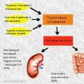 Uric acid in the blood: norms and deviations, why it increases, diet to reduce it The end product of nitrogen metabolism is