Department of Faculty Therapy. Faculty therapy
In 1966-1976 The department was headed by a brilliant teacher and clinician, Professor Anna Maksimova Tokareva. Research work was carried out on the problems of hypertension, coronary heart disease, and atherosclerosis.
In 1978-1999 The department was headed by Professor Gennady Petrovich Kuznetsov. He was one of the first in our country to study the problem of cardiomyopathies. Founder and director of the regional center for the diagnosis and treatment of cardiomyopathies. G. P. Kuznetsov is a laureate of the 1st All-Russian competition “Labor Glory of Russia” (2000), awarded the Provincial Prize (2007), the National Prize for the best doctors of Russia “For fidelity to the profession (2011), etc. by the American Bibliographical Institute (Los Angeles) is included among the outstanding personalities and awarded the “Medal of the 2000th Anniversary of Glory”.
In 1999-2009 The department was headed by Professor Vasily Vasilyevich Simerzin. He worked his way up from a nurse at the faculty therapy clinic to the head of the department. In 2002, under his leadership, the “Center for the Prevention and Treatment of Atherosclerosis and Dyslipidemia” was created. Professor V.V. Simerzin is a member of the board of the National Society for the Study of Atherosclerosis.
Department staff
From 2009 to 2014 The department was headed by Doctor of Medical Sciences, Associate Professor Mikhail Arkadyevich Kachkovsky - a member of the European Society of Cardiology, the European Society of Atherosclerosis, the All-Russian Scientific Society of Cardiologists, and the Russian Gastroenterological Association. Under his leadership, centers for the diagnosis and treatment of cardiomyopathies, the prevention and treatment of atherosclerosis and dyslipidemia continued to operate. Along with this, the gastroenterological focus of the department and clinic’s activities was actively developing.
Since 2014, the department has been headed by Oleg Veniaminovich Fatenkov, Doctor of Medical Sciences, Associate Professor, Chief Freelance Specialist in Therapy of the Ministry of Health of the Samara Region, a member of the board of the Russian National Society for the Study of Atherosclerosis. During this time, the educational process at the department was given a practically oriented orientation. Thanks to the creation of a research laboratory, innovative scientific research has begun.
The main clinical bases of the department are the Clinics of the Samara State Medical University, the Samara Regional Clinical Cardiology Dispensary, the Samara Regional Clinical Hospital named after M. I. Kalinin, city clinical hospitals No. 3, No. 6, city clinic No. 15, Medical Unit No. 2.
The department trains students, clinical interns and clinical residents in the specialty “therapy”, and conducts cycles of professional retraining and improvement according to the IPO plan.
The department takes an active part in research work. Employees of the department annually make presentations at regional and all-Russian conferences. Scientific research is being conducted in the following areas: dyslipidemia and atherosclerosis, coronary heart disease, metabolic syndrome, arterial hypertension, cardiomyopathy, pathology of the esophagus, stomach, liver, etc.
1. Fluid in the pleural cavity syndrome
2. Pleural murmur syndrome
3. Pleural air syndrome
4. Inflammatory lung tissue consolidation syndrome
Diagnosis of respiratory diseases is based on clinical, instrumental, and laboratory criteria. The set of deviations obtained when using various research methods for any pathological condition is usually called a syndrome.
1. Syndrome of fluid in the pleural cavity. A typical complaint is shortness of breath. It reflects the degree of respiratory failure due to pleural compression of the lung, which leads to a decrease in the respiratory surface in the lungs as a whole. When examining, pay attention to the protrusion and lag in the act of breathing of the corresponding half of the chest. Voice tremors and bronchotonia are weakened or absent. Percussion reveals a shortening or dullness of sound or a dull sound. On auscultation, breathing is weakened or absent.
2. Pleural murmur syndrome. Inflammation of the pleural layers can leave behind a pronounced intrapleural adhesive substrate in the form of adhesive cords, adhesions, and fibrinous pleural overlays. Such patients may have no complaints, but with severe adhesions, shortness of breath and chest pain appear during physical activity. When examining the chest, retraction and lag in the act of breathing of the affected half are noted: here you can also detect retraction of the intercostal spaces during inspiration. Voice tremors and bronchophony are weakened or absent. Percussion sound is dull. On auscultation, breathing is weakened or absent. A pleural friction rub is often heard.
3. Syndrome of air in the pleural cavity. Air may enter the pleural cavity when a subpleural cavity or abscess breaks into it. The communication of the bronchus with the pleural cavity leads to the accumulation of air in the latter, which compresses the lung. In this situation, increased pressure in the pleural cavity can lead to the closing of the hole in the pleura with pieces of damaged tissue, stopping the flow of air into the pleural cavity and the formation of a closed pneumothorax. If the connection between the bronchus and the pleural cavity is not eliminated, the pneumothorax is called an open pneumothorax.
In both cases, the main complaints are rapidly developing suffocation and chest pain. Upon examination, protrusion of the affected half of the chest and weakening of its participation in the act of breathing are determined. Voice tremors and bronchophony with closed pneumothorax are weakened or absent, with open pneumothorax they are enhanced. On percussion, tympanitis is determined in both cases. On auscultation, with a closed pneumothorax, breathing is sharply weakened or absent; with an open pneumothorax, breathing is bronchial.
In the latter case, metallic breathing may be heard as a type of bronchial breathing.
4. Inflammatory compaction syndrome of lung tissue. Compaction of lung tissue can occur not only as a result of the inflammatory process (pneumonia), when the alveoli are filled with exudate and fibrin. Compaction can occur as a result of a pulmonary infarction, when the alveoli are filled with blood, or during pulmonary edema, when edematous fluid—transudate—accumulates in the alveoli. However, inflammatory compaction of lung tissue is most common. When an entire lobe of the lung is affected, lobar or lobar pneumonia develops; one or more segments – polysegmental pneumonia; less than one segment – focal pneumonia.
Patients complain of cough, shortness of breath, and if the pleura is involved in the inflammatory process, chest pain. On examination, the affected half of the chest lags behind in the act of breathing, which is typical for lobar pneumonia. Voice tremors and bronchophony in the compaction area are increased. The percussion sound in focal pneumonia is dull (not dull), since the area of compacted lung tissue is surrounded by normal lung tissue. With lobar pneumonia, in the initial stage the sound is dull-tympanic, in the peak stage it is dull, which in the resolution stage is gradually replaced by a clear pulmonary sound.
With focal pneumonia, auscultation reveals mixed (bronchovesicular) breathing. Dry and wet rales are heard, while wet rales are characterized as sonorous, since the inflammatory compaction of the lung tissue around the bronchi promotes better conduction of the wet rales that originate in them to the surface of the chest. If the source of inflammation is deeply located, it is not possible to detect any abnormalities during physical examination. At the same time, a large focus of inflammation, located in close proximity to the visceral pleura, gives the same abnormalities during physical examination as lobar pneumonia.
In case of lobar pneumonia, auscultation on the affected side in the initial stage reveals a weakening of vesicular breathing, crepitus and pleural friction noise; at the height of the stage, bronchial breathing is heard, and there may be a pleural friction noise. In the resolution stage, bronchial breathing gradually gives way to vesicular breathing, crepitus appears, moist sonorous rales due to the penetration of liquefied exudate from the alveoli, and pleural friction noise is possible.
LECTURE 2. Clinical syndromes in respiratory diseases. Part 2
1. Lung cavity syndrome
2. Bronchospasm syndrome
3. Bronchitis syndrome
1. Lung cavity syndrome. The cavity is formed by an abscess, tuberculosis cavity, and decay of a lung tumor. A cavity formed in the lung can be detected with a diameter of at least 4 cm, communication with a bronchus, close contact with the chest wall, and a significant air content.
Patients with a destruction cavity complain of a cough with a large amount of foul-smelling yellow-green sputum. When examining the chest, a lag in the act of breathing of the affected half is detected, increased vocal tremors and bronchophony are detected, with percussion - tympanitis, with auscultation - bronchial or amphoric breathing, sonorous medium- and coarse-bubbly moist rales are heard.
2. Bronchospasm syndrome. Bronchospasm is a complex of clinical signs that occurs in the form of attacks in patients with bronchial asthma. A tendency to paroxysmal bronchospasm occurs in patients with morphologically intact bronchi and in patients with chronic bronchitis. At the moment of bronchial spasm, the patient experiences an attack of suffocation, during which exhalation is especially difficult; at the height of the attack, a cough appears with difficult to separate viscous sputum. Upon examination, the patient’s position is forced (sitting), breathing is noisy, wheezing is distant, exhalation is sharply prolonged, the veins of the neck are swollen. Accessory muscles are actively involved in the act of breathing, and diffuse cyanosis occurs. The chest is in a state of expiratory tension and has a barrel-shaped appearance. Auscultation reveals a sharply prolonged exhalation and weakening of vesicular breathing; Abundant dry whistling rales are heard in large numbers.
3. Bronchitis syndrome. With inflammation of the bronchi (bronchitis), patients complain of a cough, which at the beginning of the disease is dry, then with sputum. On examination, no deviations from the norm are detected, vocal tremors and bronchophony are not changed, and upon percussion there is a clear pulmonary sound. Auscultation - hard breathing, at the beginning of the disease dry whistling and buzzing wheezing is heard, later on moist, different-sized silent wheezing is heard, scattered throughout the lung fields.
LECTURE 3. Methods of functional diagnostics. Part 1
1. Pleural puncture
2. Sputum examination
1. Pleural puncture used to determine the nature of the pleural fluid in order to clarify the diagnosis, to remove fluid from the pleural cavity and subsequently introduce drugs into it.
The puncture is usually performed along the posterior axillary line in the seventh or eighth intercostal space along the upper edge of the rib. For diagnostic purposes, 50-150 ml of liquid is removed, which is sent for cytological and bacteriological examination. For therapeutic purposes, when a large amount of fluid accumulates in the pleural cavity, 800-1200 ml are initially evacuated. Removal of a larger amount of fluid leads to a rapid displacement of the mediastinal organs to the affected side and may be accompanied by collapse. Pleural fluid can be of inflammatory (exudate) or non-inflammatory (transudate) origin. When analyzed in a clinical laboratory, the specific gravity, the amount of protein contained, red blood cells, leukocytes, mesothelial and atypical cells are determined. The specific gravity of the inflammatory fluid is 1.015 or higher, the protein content is more than 2–3%, the Rivald test is positive. The specific gravity of the transudate is less than 1.015, the amount of protein is less than 2%, the Rivald test is negative.
2. Sputum examination. Sputum is a pathological discharge of the broncho-alveolar tract, expelled during coughing and expectoration. Sputum may contain mucus, serous fluid, blood and respiratory tract cells, tissue decay elements, crystals, microorganisms, protozoa, helminths and their eggs. Examination of sputum helps to establish the nature of the pathological process in the respiratory organs, and in some cases determine its etiology.
It is better to take sputum for examination in the morning, if possible before meals and after rinsing the mouth. However, to detect tuberculosis microbacteria, sputum, if the patient produces little of it, must be collected within 1–2 days.
The daily amount of sputum ranges from 1 to 1000 ml or more. Mucous sputum is usually colorless or slightly whitish, viscous; separated, for example, in acute bronchitis. Serous sputum is also colorless, liquid, foamy. Mucopurulent sputum – yellow or greenish in color, viscous; formed in chronic bronchitis and tuberculosis. Purulent sputum is homogeneous, semi-liquid, greenish-yellow, characteristic of an abscess when it ruptures. Bloody sputum can be either pure blood in pulmonary hemorrhages or mixed.
Sputum often has no odor. Kurshman spirals can be found in sputum in the form of small, dense, tortuous whitish threads; fibrin clots (whitish and reddish tree-like branched formations found in fibrinous bronchitis, occasionally in pneumonia); small dense, greenish-yellow lumps consisting of calcified elastic fibers, crystals, cholesterol, containing Mycobacterium tuberculosis; lime grains found during the disintegration of old tuberculosis lesions; drusen of actinomycetes in the form of small yellowish grains; necrotic pieces of lung tissue; leftover food.
Microscopic examination of sputum is carried out in both native and stained preparations. For the first, pathological lumps are selected from the material poured into a Petri dish and transferred to a glass slide in such an amount that when covered with a cover glass, a thin translucent preparation is formed. The latter is viewed first at low magnification for initial orientation and search for Kurshman spirals, and then at high magnification to differentiate the shaped elements. Courshmann spirals are strands of mucus, consisting of a central dense axial thread and a spiral-shaped “matia” enveloping it, in which leukocytes (often eosinophilic) Charcot-Leyden crystals are interspersed. Kurshman spirals appear in sputum during bronchospasm, most often with bronchial asthma, less often with pneumonia.
With high magnification, leukocytes can be detected in the native preparation, a small number of which is present in any sputum, and a large number in inflammatory and, in particular, suppurative processes; Eosinophilic leukocytes can be distinguished in a native preparation by their uniform, large, shiny granularity, but they are easier to recognize when stained. Red blood cells appear during the destruction of lung tissue, pneumonia, stagnation in the pulmonary circulation, or pulmonary infarction. Flat epithelium enters the sputum mainly from the oral cavity and has no diagnostic value. Columnar ciliated epithelium is present in small quantities in every sputum, and in large quantities in cases of damage to the respiratory tract (bronchitis, bronchial asthma). Alveolar macrophages are large cells (2-3 times more leukocytes) of reticuloendothelial origin. Their cytoplasm contains abundant inclusions. The latter can be colorless (myelin grains), black from coal particles (dust cells) or yellow-brown from hemosiderin (“heart defect cells”, siderophages). Alveolar macrophages are present in small quantities in each sputum; they are more numerous in inflammatory diseases; cells of heart defects occur when red blood cells enter the cavity of the alveoli; with stagnation in the pulmonary circulation, especially with mitral stenosis; for pulmonary infarction, hemorrhage, and pneumonia.
LECTURE 4. Methods of functional diagnostics. Part 2
1. X-ray examination
2. Endoscopic examination
1. To examine the respiratory organs, fluoroscopy, radiography, bronchography and tomography of the lungs are used.
Fluoroscopy allows you to visually determine changes in the transparency of the lung tissue, detect foci of compaction or cavities in it, identify the presence of fluid or air in the pleural cavity, as well as other pathological changes.
Radiography is used to record and document changes in the respiratory organs detected during fluoroscopy on x-ray film. In pathological processes in the lungs, leading to loss of airiness and compaction of the lung tissue (pneumonia, pulmonary infarction, tuberculosis, etc.), the corresponding areas of the lungs on the film have a paler image compared to normal lung tissue. The cavity in the lung, containing air and surrounded by an inflammatory shaft, has the appearance of a dark oval-shaped spot, surrounded by a paler shadow than the shadow of the lung tissue. Fluid in the pleural cavity, which transmits fewer x-rays than lung tissue, produces a shadow on negative x-ray film that is paler than that of lung tissue.
The X-ray method allows you to determine not only the amount of fluid in the pleural cavity, but also its nature. If there is exudate in the pleural cavity, the level of its contact with the lung has an oblique line, gradually directed upward and laterally from the midclavicular line; when transudate accumulates in the pleural cavity, the level is located more horizontally.
Tomography allows for layer-by-layer X-ray examination of the lungs. It is used to diagnose tumors of the bronchi and lungs, small infiltrates, cavities and cavities located at various depths.
Bronchography is used to study the bronchi. After preliminary anesthesia, the patient is injected into the lumen of the bronchi with a contrast agent that blocks x-rays, then an x-ray of the lungs is taken and a clear image of the bronchial tree is obtained on the x-ray. This method allows you to diagnose bronchial dilatation (bronchiectasis), abscesses and lung caverns, narrowing of the lumen of large bronchi by a tumor or foreign body.
Fluorography is also a type of x-ray examination of the lungs. It is used for mass preventive examination of the population.
2. Endoscopic research methods include broncho- and thoracoscopy.
Bronchoscopy allows you to examine the mucous membrane of the trachea and bronchi of the first, second and third order. It is performed with a special device - a bronchoscope, which is equipped with special forceps for biopsy, removal of foreign bodies, removal of polyps, a photo attachment, etc. Before inserting the bronchoscope, anesthesia is performed with a 1-3% solution of dicaine of the mucous membrane of the upper respiratory tract, then the bronchoscope is inserted through mouth and glottis into the trachea. The doctor examines the mucous membrane of the trachea and bronchi.
For histological and cytological studies, a piece of tissue from a suspicious area can be removed (biopsy) using special forceps. Bronchoscopy is used to diagnose erosions, ulcers of the bronchial mucosa and tumors of the bronchial wall, remove foreign bodies, remove bronchial polyps, treat bronchiectasis and centrally located lung abscesses. In these cases, purulent sputum is first sucked out through a bronchoscope, and then antibiotics are injected into the bronchial lumen or cavity.
LECTURE 5. Pneumonia. Etiology, pathogenesis, classification
1. Definition and etiology
2. Pathogenesis
3. Classification of pneumonia
1. Pneumonia – acute infectious exudative inflammation of the respiratory parts of the lungs, which is caused by microorganisms of various natures and covers the distal parts of the respiratory tract.
Etiology. The most common causative agents of community-acquired pneumonia are pneumococci (30–40%), mycoplasma (up to 20%) and viruses (10%). In the case of hospital-acquired pneumonia, the causative agents are usually Pseudomonas aeruginosa, Proteus, Legionella, Aspirgillus, Mycoplasma and Pneumocystis. In aspiration pneumonia, the causative agents most often are associations of gram-positive and gram-negative bacteria with anaerobic microorganisms. Such pneumonia occurs in diseases of the gastrointestinal tract, nervous system, and in patients with multiple injuries. In adolescents, the most common cause of pneumonia is Mykoplasma pneumoniae.
2. Pathogenesis. The disease occurs under conditions of superinfection or high virulence of the microorganism, on the one hand, and decreased immunity, on the other. Microorganisms penetrate into the lung tissue in one of four ways: by inhalation with air; aspiration from the oro- or nasopharynx; hematogenous spread of their distant source of infection; lymphogenous (from neighboring organs) when the chest is injured. The most typical route of infection is inhalation. A certain role in the pathogenesis of pneumonia is played by violations of the cellular mechanism of anti-infective defense, neurotrophic disorder of the bronchi and lungs associated with the effect of bacterial flora on the interoreceptor apparatus of the respiratory tract.
Predisposing factors for community-acquired pneumonia are smoking, stress, hypothermia, physical fatigue and emotional stress, prolonged exposure to a working air conditioner, impaired consciousness of any etiology, epilepsy with frequent seizures, artificial ventilation, alcohol intoxication, immunodeficiency states (primary or secondary). These factors, suppressing local protective mechanisms and disrupting pulmonary circulation, lead to damage to the bronchioles and alveoli.
3. Classification of pneumonia. According to the international classification (European Respiratory Society, 1995), pneumonia is distinguished:
1) community-acquired (primary);
2) nosocomial (nosocomial), occurring 48–72 hours after hospitalization for other diseases;
3) in persons with severe immune defects;
4) atypical.
The previously widespread classification of pneumonia according to pathomorphological characteristics into lobar and focal provides relatively little information for choosing optimal etiotropic therapy. From a practical point of view, it should be considered more rational to distinguish between two main classes of pneumonia – “community-acquired” and “nosocomial-acquired”. Each class is characterized not only by its place of origin, but also has its own significant features (epidemiological, clinical, radiological, etc.), and most importantly, a certain spectrum of pathogens.
From these positions, pneumonia is distinguished that occurs in closely interacting teams, the features of which are as follows:
– occur, as a rule, in previously healthy people in the absence of background pathology;
– the disease is most common in the winter season, which is explained by the high frequency of infections with the influenza A virus, respiratory infection virus, certain epidemiological situations (viral epidemics, outbreaks of mycoplasma infection, Q-fever, etc.);
– risk factors are contact with animals, birds (ornithosis, psittacosis), contact with air conditioners (legionella pneumonia);
– main pathogens: pneumococcus, mycoplasma, legionella, chlamydia, various viruses, hemophilus influenzae.
Nosocomial (hospital-acquired) pneumonia is characterized by the following features:
– occur after two or more days of hospital stay in the absence of clinical and radiological signs of pulmonary damage during hospitalization;
– are one of the forms of nosocomial (hospital) infections and occupy third place after urinary tract infections and wound infections;
– mortality from hospital pneumonia is about 20% – risk factors are the very fact that patients are in intensive care wards, intensive care units, the presence of artificial ventilation, tracheostomy, bronchoscopic examinations, the postoperative period (especially after thoracoabdominal operations), massive antibiotic therapy, septic conditions ;
– the main pathogens are gram-negative microorganisms, staphylococcus.
Pneumonia is classified according to severity: mild, moderate, severe, and extremely severe.
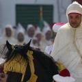 King Mohammed VI of Morocco divorced Princess Lalla Salma State of the country at the beginning of his reign
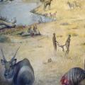 The emergence of life on Earth
 Notes on the stones. Lev Shlosberg. Notes on stones Main works of Vsevolod Petrovich Smirnov