Which organisms first developed a nervous system? What animal first developed a nervous system? General characteristics of the nervous system
In evolution, the nervous system has undergone several stages of development, which became turning points in the qualitative organization of its activities. These stages differ in the number and types of neuronal formations, synapses, signs of their functional specialization, and in the formation of groups of neurons interconnected by common functions. There are three main stages of the structural organization of the nervous system: diffuse, nodular, tubular.
Diffuse The nervous system is the most ancient, found in coelenterates (hydra). Such a nervous system is characterized by a multiplicity of connections between neighboring elements, which allows excitation to freely spread throughout the nervous network in all directions.
This type of nervous system provides wide interchangeability and thereby greater reliability of functioning, but these reactions are imprecise and vague.
Nodal the type of nervous system is typical for worms, mollusks, and crustaceans.
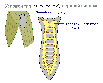
It is characterized by the fact that the connections of nerve cells are organized in a certain way, excitation passes along strictly defined paths. This organization of the nervous system turns out to be more vulnerable. Damage to one node causes dysfunction of the entire organism as a whole, but its qualities are faster and more accurate.
Tubular The nervous system is characteristic of chordates; it includes features of diffuse and nodular types. The nervous system of higher animals took all the best: high reliability of the diffuse type, accuracy, locality, speed of organization of nodal type reactions.
The leading role of the nervous system
At the first stage of the development of the world of living beings, interaction between the simplest organisms was carried out through the aquatic environment of the primitive ocean, into which the chemical substances released by them entered. The first oldest form of interaction between the cells of a multicellular organism is chemical interaction through metabolic products entering the body fluids. Such metabolic products, or metabolites, are the breakdown products of proteins, carbon dioxide, etc. This is the humoral transmission of influences, the humoral mechanism of correlation, or connections between organs.
The humoral connection is characterized by the following features:
- lack of an exact address to which a chemical substance entering the blood or other body fluids is sent;
- the chemical spreads slowly;
- the chemical acts in minute quantities and is usually quickly broken down or eliminated from the body.
Humoral connections are common to both the animal and plant worlds. At a certain stage of development of the animal world, in connection with the appearance of the nervous system, a new, nervous form of connections and regulation is formed, which qualitatively distinguishes the animal world from the plant world. The higher the development of an animal’s organism, the greater the role played by the interaction of organs through the nervous system, which is designated as reflex. In higher living organisms, the nervous system regulates humoral connections. Unlike the humoral connection, the nervous connection has a precise direction to a specific organ and even a group of cells; communication is carried out hundreds of times faster than the speed of distribution of chemicals. The transition from a humoral connection to a nervous connection was not accompanied by the destruction of the humoral connection between the cells of the body, but by the subordination of nervous connections and the emergence of neurohumoral connections.
At the next stage of development of living beings, special organs appear - glands, in which hormones are produced, formed from food substances entering the body. The main function of the nervous system is both to regulate the activity of individual organs among themselves, and in the interaction of the body as a whole with its external environment. Any impact of the external environment on the body appears, first of all, on receptors (sensory organs) and is carried out through changes caused by the external environment and the nervous system. As the nervous system develops, its highest department—the cerebral hemispheres—becomes “the manager and distributor of all the activities of the body.”
Structure of the nervous system
The nervous system is formed by nervous tissue, which consists of a huge amount neurons- a nerve cell with processes.

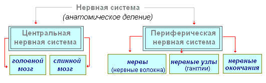
The nervous system is conventionally divided into central and peripheral.

central nervous system includes the brain and spinal cord, and peripheral nervous system- nerves extending from them.
The brain and spinal cord are a collection of neurons. In a cross section of the brain, white and gray matter are distinguished. Gray matter consists of nerve cells, and white matter consists of nerve fibers, which are processes of nerve cells. In different parts of the central nervous system, the location of white and gray matter is different. In the spinal cord, gray matter is located inside, and white matter is on the outside; in the brain (cerebral hemispheres, cerebellum), on the contrary, gray matter is on the outside, white matter is on the inside. In various parts of the brain there are separate clusters of nerve cells (gray matter) located inside the white matter - kernels. Clusters of nerve cells are also located outside the central nervous system. They're called nodes and belong to the peripheral nervous system.
Reflex activity of the nervous system
The main form of activity of the nervous system is the reflex. Reflex- the body’s reaction to changes in the internal or external environment, carried out with the participation of the central nervous system in response to irritation of receptors.
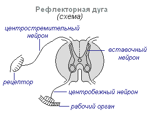
With any irritation, excitation from the receptors is transmitted along centripetal nerve fibers to the central nervous system, from where, through the interneuron along centrifugal fibers, it goes to the periphery to one or another organ, the activity of which changes. This entire path through the central nervous system to the working organ is called reflex arc usually formed by three neurons: sensory, intercalary and motor. A reflex is a complex act in which a significantly larger number of neurons take part. Excitation, entering the central nervous system, spreads to many parts of the spinal cord and reaches the brain. As a result of the interaction of many neurons, the body responds to irritation.
Spinal cord
Spinal cord- a cord about 45 cm long, 1 cm in diameter, located in the spinal canal, covered with three meninges: dura, arachnoid and soft (vascular).

Spinal cord is located in the spinal canal and is a cord that at the top passes into the medulla oblongata and at the bottom ends at the level of the second lumbar vertebra. The spinal cord consists of gray matter containing nerve cells and white matter consisting of nerve fibers. Gray matter is located inside the spinal cord and is surrounded on all sides by white matter.
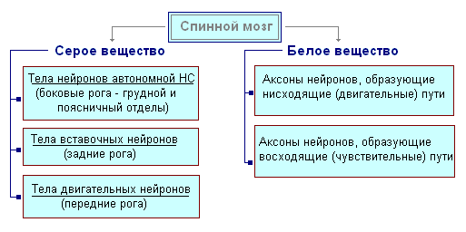
In a cross section, the gray matter resembles the letter H. It distinguishes the anterior and posterior horns, as well as the connecting crossbar, in the center of which there is a narrow canal of the spinal cord containing cerebrospinal fluid. In the thoracic region there are lateral horns. They contain the bodies of neurons that innervate internal organs. The white matter of the spinal cord is formed by nerve processes. Short processes connect sections of the spinal cord, and long ones make up the conductive apparatus of bilateral connections with the brain.

The spinal cord has two thickenings - cervical and lumbar, from which nerves extend to the upper and lower extremities. 31 pairs of spinal nerves arise from the spinal cord. Each nerve begins from the spinal cord with two roots - anterior and posterior. Posterior roots - sensitive consist of processes of centripetal neurons. Their bodies are located in the spinal ganglia. Anterior roots - motor- are processes of centrifugal neurons located in the gray matter of the spinal cord. As a result of the fusion of the anterior and posterior roots, a mixed spinal nerve is formed. The spinal cord contains centers that regulate the simplest reflex acts. The main functions of the spinal cord are reflex activity and conduction of excitation.
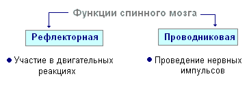
The human spinal cord contains reflex centers for the muscles of the upper and lower extremities, sweating and urination. The function of excitation is that impulses from the brain to all areas of the body and back pass through the spinal cord. Centrifugal impulses from organs (skin, muscles) are transmitted through ascending pathways to the brain. Along descending pathways, centrifugal impulses are transmitted from the brain to the spinal cord, then to the periphery, to the organs. When the pathways are damaged, there is a loss of sensitivity in various parts of the body, a violation of voluntary muscle contractions and the ability to move.
Evolution of the vertebrate brain
The formation of the central nervous system in the form of a neural tube first appears in chordates. U lower chordates the neural tube persists throughout life, higher- vertebrates - in the embryonic stage, a neural plate is laid down on the dorsal side, which sinks under the skin and curls up into a tube. In the embryonic stage of development, the neural tube forms three swellings in the anterior part - three brain vesicles, from which parts of the brain develop: the anterior vesicle gives the forebrain and diencephalon, the middle vesicle turns into the midbrain, the posterior vesicle forms the cerebellum and medulla oblongata. These five brain regions are characteristic of all vertebrates.
For lower vertebrates- fish and amphibians - characterized by a predominance of the midbrain over other parts. U amphibians The forebrain enlarges somewhat and a thin layer of nerve cells forms in the roof of the hemispheres - the primary medullary vault, the ancient cortex. U reptiles The forebrain increases significantly due to accumulations of nerve cells. Most of the roof of the hemispheres is occupied by the ancient cortex. For the first time in reptiles, the rudiment of a new cortex appears. The hemispheres of the forebrain creep onto other parts, as a result of which a bend is formed in the region of the diencephalon. Beginning with ancient reptiles, the cerebral hemispheres became the largest part of the brain.
In the structure of the brain birds and reptiles much in common. On the roof of the brain is the primary cortex, the midbrain is well developed. However, in birds, compared to reptiles, the total brain mass and the relative size of the forebrain increase. The cerebellum is large and has a folded structure. U mammals the forebrain reaches its greatest size and complexity. Most of the brain matter is made up of the neocortex, which serves as the center of higher nervous activity. The intermediate and middle parts of the brain in mammals are small. The expanding hemispheres of the forebrain cover them and crush them under themselves. Some mammals have a smooth brain without grooves or convolutions, but most mammals have grooves and convolutions in the cerebral cortex. The appearance of grooves and convolutions occurs due to the growth of the brain with limited dimensions of the skull. Further growth of the cortex leads to the appearance of folding in the form of grooves and convolutions.
Brain
If the spinal cord in all vertebrates is developed more or less equally, then the brain differs significantly in size and complexity of structure in different animals. The forebrain undergoes particularly dramatic changes during evolution. In lower vertebrates, the forebrain is poorly developed. In fish, it is represented by the olfactory lobes and nuclei of gray matter in the thickness of the brain. The intensive development of the forebrain is associated with the emergence of animals onto land. It differentiates into the diencephalon and two symmetrical hemispheres, which are called telencephalon. Gray matter on the surface of the forebrain (cortex) first appears in reptiles, developing further in birds and especially in mammals. Truly large forebrain hemispheres become only in birds and mammals. In the latter, they cover almost all other parts of the brain.
The brain is located in the cranial cavity. It includes the brainstem and telencephalon (cerebral cortex).

Brain stem consists of the medulla oblongata, pons, midbrain and diencephalon.
Medulla is a direct continuation of the spinal cord and, expanding, passes into the hindbrain. It basically retains the shape and structure of the spinal cord. In the thickness of the medulla oblongata there are accumulations of gray matter - the nuclei of the cranial nerves. The rear axle includes cerebellum and pons. The cerebellum is located above the medulla oblongata and has a complex structure. On the surface of the cerebellar hemispheres, gray matter forms the cortex, and inside the cerebellum - its nuclei. Like the spinal medulla oblongata, it performs two functions: reflex and conductive. However, the reflexes of the medulla oblongata are more complex. This is reflected in its importance in the regulation of cardiac activity, the condition of blood vessels, respiration, and sweating. The centers of all these functions are located in the medulla oblongata. Here are the centers for chewing, sucking, swallowing, saliva and gastric juice. Despite its small size (2.5–3 cm), the medulla oblongata is a vital part of the central nervous system. Damage to it can cause death due to cessation of breathing and heart activity. The conductor function of the medulla oblongata and the pons is to transmit impulses from the spinal cord to the brain and back.
IN midbrain the primary (subcortical) centers of vision and hearing are located, which carry out reflexive orienting reactions to light and sound stimulation. These reactions are expressed in various movements of the torso, head and eyes towards the stimuli. The midbrain consists of the cerebral peduncles and quadrigeminalis. The midbrain regulates and distributes the tone (tension) of skeletal muscles.
Diencephalon consists of two departments - thalamus and hypothalamus, each of which consists of a large number of nuclei of the visual thalamus and subthalamic region. Through the visual thalamus, centripetal impulses are transmitted to the cerebral cortex from all receptors of the body. Not a single centripetal impulse, no matter where it comes from, can pass to the cortex, bypassing the visual hillocks. Thus, through the diencephalon, all receptors communicate with the cerebral cortex. In the subtubercular region there are centers that influence metabolism, thermoregulation and endocrine glands.
Cerebellum located behind the medulla oblongata. It consists of gray and white matter. However, unlike the spinal cord and brainstem, the gray matter - the cortex - is located on the surface of the cerebellum, and the white matter is located inside, under the cortex. The cerebellum coordinates movements, makes them clear and smooth, plays an important role in maintaining the balance of the body in space, and also influences muscle tone. When the cerebellum is damaged, a person experiences a decrease in muscle tone, movement disorders and changes in gait, speech slows down, etc. However, after some time, movement and muscle tone are restored due to the fact that the intact parts of the central nervous system take over the functions of the cerebellum.
Large hemispheres- the largest and most developed part of the brain. In humans, they form the bulk of the brain and are covered with cortex over their entire surface. Gray matter covers the outside of the hemispheres and forms the cerebral cortex. The human cerebral cortex has a thickness of 2 to 4 mm and is composed of 6–8 layers formed by 14–16 billion cells, different in shape, size and functions. Under the cortex is a white substance. It consists of nerve fibers connecting the cortex with the lower parts of the central nervous system and the individual lobes of the hemispheres with each other.
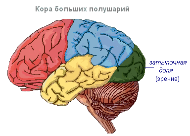
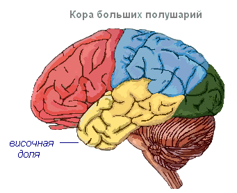
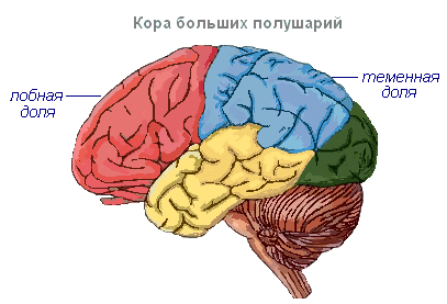
The cerebral cortex has convolutions separated by grooves, which significantly increase its surface. The three deepest grooves divide the hemispheres into lobes. Each hemisphere has four lobes: frontal, parietal, temporal, occipital. Excitation of different receptors enters the corresponding perceptive areas of the cortex, called zones, and from here they are transmitted to a specific organ, prompting it to action. The following zones are distinguished in the cortex. Auditory zone located in the temporal lobe, receives impulses from auditory receptors.

Visual area lies in the occipital region. Impulses from the eye receptors arrive here.
Olfactory zone located on the inner surface of the temporal lobe and is associated with receptors in the nasal cavity.
Sensory-motor the zone is located in the frontal and parietal lobes. This zone contains the main centers of movement of the legs, torso, arms, neck, tongue and lips. This is also where the center of speech lies.
The cerebral hemispheres are the highest division of the central nervous system, controlling the functioning of all organs in mammals. The importance of the cerebral hemispheres in humans also lies in the fact that they represent the material basis of mental activity. I.P. Pavlov showed that mental activity is based on physiological processes occurring in the cerebral cortex. Thinking is associated with the activity of the entire cerebral cortex, and not just with the function of its individual areas.
| Brain department | Functions | |
| Medulla | Conductor | Connection between the spinal and overlying parts of the brain. |
| Reflex | Regulation of the activity of the respiratory, cardiovascular, digestive systems:
|
|
| Pons | Conductor | Connects the cerebellar hemispheres to each other and to the cerebral cortex. |
| Cerebellum | Coordination | Coordination of voluntary movements and maintaining body position in space. Regulation of muscle tone and balance |
| Midbrain | Conductor | Approximate reflexes to visual and sound stimuli ( turns the head and body). |
| Reflex |
|
|
| Diencephalon | thalamus
hypothalamus
|
|
Cerebral cortex
Surface cerebral cortex in humans it is about 1500 cm 2, which is many times greater than the inner surface of the skull. This large surface of the cortex was formed due to the development of a large number of grooves and convolutions, as a result of which most of the cortex (about 70%) is concentrated in the grooves. The largest grooves of the cerebral hemispheres are central, which runs across both hemispheres, and temporal, separating the temporal lobe from the rest. The cerebral cortex, despite its small thickness (1.5–3 mm), has a very complex structure. It has six main layers, which differ in the structure, shape and size of neurons and connections. The cortex contains the centers of all sensory (receptor) systems, representatives of all organs and parts of the body. In this regard, centripetal nerve impulses from all internal organs or parts of the body approach the cortex, and it can control their work. Through the cerebral cortex, conditioned reflexes are closed, through which the body constantly, throughout life, very accurately adapts to the changing conditions of existence, to the environment.
The nervous system in a living organism is represented by a network of communications that ensure its connection with the outside world and its own processes. Its basic element is a neuron - a cell with processes (axons and dendrites) that transmit information electrically and chemically.
Purpose of nervous regulation
For the first time, the nervous system appeared in living organisms due to the need for more effective interaction with the environment. The development of a simple network for transmitting impulses helped not only to perceive signals from the outside. Thanks to it, it became possible to organize one’s own life processes for more successful functioning.
During evolution, the structure of the nervous system became more complex: its task became not only the formation of an adequate response to external influences, but also the organization of its own behavior. I. P. Pavlov called this method of functioning
Interaction with the unicellular environment
The nervous system first appeared in organisms consisting of more than one cell, as it transmits signals between neurons that form a network. But already in protozoa one can observe the ability to respond to external stimuli provided by intracellular processes.
The nervous system of multicellular organisms is qualitatively different from a similar formation in protozoa. The latter locate the entire system of connections within the metabolism of a single cell. The ciliates “learn” about various processes that occur externally or internally due to changes in the composition of the protoplasm and the activity of some other structures. Multicellular living beings have a system built from functional units, each of which is endowed with its own metabolic processes.
Thus, for the first time, a nervous system appears in someone who has not one, but several cells, that is, the prototype is the conduction of impulses in protozoa. At their level of vital activity, the protoplasm produces structures that conduct impulses. Similarly, in more complex living beings, this function is performed by individual
Features of the nervous system of coelenterates
Multicellular animals living in colonies do not share functions among themselves, and they do not yet have a nervous network. It occurs at the stage when various functions in a multicellular organism are differentiated.

For the first time, the nervous system appears in hydra and other coelenterates. It is a network that conducts non-targeted signals. The structure has not yet been formed; it is diffusely distributed throughout the body of the coelenterate. Ganglion cells and their Nisslev substance are not fully formed. This is the simplest version of the nervous system.
The type of animal motor activity is determined by the diffuse network-like nervous system. Hydra performs peristaltic movements, since it does not have special body parts for movement and other movements. For motor activity, it requires continuous connection of the contractile elements, and it requires that the bulk of the conducting cells be located in the contractile part. In which animal does the nervous system first appear in the form of a diffuse network? Those who are the founders of the human regulatory system. Proof of this is the fact that gastrulation is present in the development of animal embryos.
Features of the nervous system of helminths
Subsequent improvement of nervous regulation was associated with the development of bilateral symmetry instead of radial and the formation of clusters of neurons in various parts of the body.

In the form of cords, the nervous system first appears in 1 At this stage, it is represented by paired head fibers and formed fibers extending from them. Compared to coelenterates, this system is much more complex. In helminths, groups of nerve cells are found in the form of nodes and ganglia. The prototype of the brain is a ganglion in the front of the body that performs regulatory functions. It is called the cerebral ganglion. From it, two nerve trunks run along the entire body, connected by jumpers.

All components of the system are not located outside, but are immersed in the parenchyma and are thereby protected from injury. For the first time, the nervous system appears in flatworms along with the simplest sense organs: touch, vision and a sense of balance.
Features of the nervous system of nematodes
The next stage of development is the formation of a ring formation near the pharynx and several long fibers extending from it. With such characteristics, the nervous system first appears in the Peripharyngeal ring, which is a single circular ganglion and performs the functions of the basic organ of perception. The ventral cord and dorsal nerve are connected to it.
The nerve trunks of nematodes are located intraepithelially, that is, in the hypodermal ridges. The role of perception organs is played by sensilla - setae, papillae, supplementary organs, amphids and phasmids. All of them are endowed with mixed sensitivity.

The most complex perception organs of nematodes are amphids. They are paired, can be different in shape and are located in the front. Their main task is to recognize chemical agents located far from the body. Some roundworms also have receptors that perceive internal and external mechanical influences. They are called metanemes.
Features of the ringlet nervous system
The formation of ganglia in the nervous system further develops in annelids. In most of them, ganglionization of the abdominal trunks occurs in such a way that each segment of the worm has a pair of nerve nodes that are connected by fibers to neighboring segments. have a ventral nerve cord formed by the medullary ganglion and a pair of cords coming from it. They stretch along the abdominal plane. Perceiving elements are located in front and are represented by the simplest eyes, olfactory cells, ciliary fossae and locators. With paired nodes, the nervous system first appeared in annelids, but later it develops in arthropods. They have an enlargement of the ganglia in the head and a combination of nodes in the body.
Elements of the diffuse network in the human nervous system
The peak of the evolutionary development of the nervous system is the appearance of the brain and spinal cord in humans. However, even in the presence of such complex structures, the original diffuse organization is preserved. This network entangles every cell of the body: skin, blood vessels, etc. But with such characteristics, for the first time, a nervous system appears in someone who did not even have the opportunity to differentially perceive the environment.

Thanks to these “residual” structural units, a person has the opportunity to feel various influences even in microscopic areas. The body can react to the appearance of the smallest foreign agent by developing protective reactions. The presence of a diffuse network in the human nervous system is confirmed by laboratory research methods based on the introduction of a dye.
General line of development of the nervous system during evolution
The evolutionary processes of the nervous system took place in three stages:
- diffuse network;
- gangilia;
- spinal cord and brain.

The structure and functioning of the CNS is very different from earlier types. Its sympathetic division contains ganglionic and reticular elements. In its phylogenetic development, the nervous system became increasingly fragmented and differentiated. The ganglionic stage of development differed from the reticular stage in the presence of neurons still located above the conduction system.
Any living organism is essentially a monolith, consisting of various organs and their systems, which constantly and continuously interact with each other and with the external environment. The nervous system first appeared in coelenterates; it was a diffuse network that ensured the elementary conduction of impulses.
LECTURE ON THE TOPIC: HUMAN NERVOUS SYSTEM
Nervous system is a system that regulates the activities of all human organs and systems. This system determines: 1) the functional unity of all human organs and systems; 2) the connection of the whole organism with the environment.
From the point of view of maintaining homeostasis, the nervous system ensures: maintaining the parameters of the internal environment at a given level; inclusion of behavioral responses; adaptation to new conditions if they persist for a long time.
Neuron(nerve cell) - the main structural and functional element of the nervous system; Humans have more than one hundred billion neurons. A neuron consists of a body and processes, usually one long process - an axon and several short branched processes - dendrites. Along dendrites, impulses follow to the cell body, along an axon - from the cell body to other neurons, muscles or glands. Thanks to the processes, neurons contact each other and form neural networks and circles through which nerve impulses circulate.
A neuron is a functional unit of the nervous system. Neurons are susceptible to stimulation, that is, they are capable of being excited and transmitting electrical impulses from receptors to effectors. Based on the direction of impulse transmission, afferent neurons (sensory neurons), efferent neurons (motor neurons) and interneurons are distinguished.
Nervous tissue is called excitable tissue. In response to some impact, a process of excitation arises and spreads in it - rapid recharging of cell membranes. The emergence and propagation of excitation (nerve impulse) is the main way the nervous system carries out its control function.
The main prerequisites for the occurrence of excitation in cells: the existence of an electrical signal on the membrane in a resting state - the resting membrane potential (RMP);
the ability to change the potential by changing the permeability of the membrane for certain ions.
The cell membrane is a semi-permeable biological membrane, it has channels that allow potassium ions to pass through, but there are no channels for intracellular anions, which are retained at the inner surface of the membrane, creating a negative charge of the membrane from the inside, this is the resting membrane potential, which averages - – 70 millivolts (mV). There are 20-50 times more potassium ions in the cell than outside, this is maintained throughout life with the help of membrane pumps (large protein molecules capable of transporting potassium ions from the extracellular environment to the inside). The MPP value is determined by the transfer of potassium ions in two directions:
1. from the outside into the cell under the action of pumps (with a large expenditure of energy);
2. from the cell to the outside by diffusion through membrane channels (without energy consumption).
In the process of excitation, the main role is played by sodium ions, which are always 8-10 times more abundant outside the cell than inside. Sodium channels are closed when the cell is at rest; in order to open them, it is necessary to act on the cell with an adequate stimulus. If the stimulation threshold is reached, the sodium channels open and sodium enters the cell. In thousandths of a second, the membrane charge will first disappear and then change to the opposite - this is the first phase of the action potential (AP) - depolarization. The channels close - the peak of the curve, then the charge is restored on both sides of the membrane (due to potassium channels) - the repolarization stage. The excitation stops and while the cell is at rest, the pumps exchange the sodium that entered the cell for potassium, which left the cell.

An PD evoked at any point in a nerve fiber itself becomes an irritant for neighboring sections of the membrane, causing AP in them, which in turn excite more and more sections of the membrane, thus spreading throughout the entire cell. In fibers covered with myelin, APs will occur only in areas free of myelin. Therefore, the speed of signal propagation increases.


The transfer of excitation from cell to another occurs through a chemical synapse, which is represented by the point of contact of two cells. The synapse is formed by presynaptic and postsynaptic membranes and the synaptic cleft between them. Excitation in the cell resulting from AP reaches the area of the presynaptic membrane where synaptic vesicles are located, from which a special substance, the transmitter, is released. The transmitter entering the gap moves to the postsynaptic membrane and binds to it. Pores open in the membrane for ions, they move into the cell and the process of excitation occurs

Thus, in the cell, the electrical signal is converted into a chemical one, and the chemical signal again into an electrical one. Signal transmission in a synapse occurs more slowly than in a nerve cell, and is also one-sided, since the transmitter is released only through the presynaptic membrane, and can only bind to receptors of the postsynaptic membrane, and not vice versa.
Mediators can cause not only excitation but also inhibition in cells. In this case, pores open on the membrane for ions that strengthen the negative charge that existed on the membrane at rest. One cell can have many synaptic contacts. An example of a mediator between a neuron and a skeletal muscle fiber is acetylcholine.
The nervous system is divided into central nervous system and peripheral nervous system.
In the central nervous system, a distinction is made between the brain, where the main nerve centers and the spinal cord are concentrated, and here there are lower-level centers and pathways to peripheral organs.
Peripheral section - nerves, nerve ganglia, ganglia and plexuses.
The main mechanism of activity of the nervous system is reflex. A reflex is any response of the body to a change in the external or internal environment, which is carried out with the participation of the central nervous system in response to irritation of receptors. The structural basis of the reflex is the reflex arc. It includes five consecutive links:
1 - Receptor - a signaling device that perceives influence;
2 - Afferent neuron – brings a signal from the receptor to the nerve center;
3 - Interneuron – central part of the arc;
4 - Efferent neuron - the signal comes from the central nervous system to the executive structure;
5 - Effector - a muscle or gland performing a certain type of activity

Brain consists of clusters of nerve cell bodies, nerve tracts and blood vessels. Nerve tracts form the white matter of the brain and consist of bundles of nerve fibers that conduct impulses to or from various parts of the gray matter of the brain - nuclei or centers. Pathways connect various nuclei, as well as the brain and spinal cord.
Functionally, the brain can be divided into several sections: the forebrain (consisting of the telencephalon and diencephalon), the midbrain, the hindbrain (consisting of the cerebellum and the pons) and the medulla oblongata. The medulla oblongata, pons, and midbrain are collectively called the brainstem.

Spinal cord located in the spinal canal, reliably protecting it from mechanical damage.
The spinal cord has a segmental structure. Two pairs of anterior and posterior roots extend from each segment, which corresponds to one vertebra. There are 31 pairs of nerves in total.
The dorsal roots are formed by sensory (afferent) neurons, their bodies are located in the ganglia, and the axons enter the spinal cord.
The anterior roots are formed by the axons of efferent (motor) neurons, the bodies of which lie in the spinal cord.
The spinal cord is conventionally divided into four sections - cervical, thoracic, lumbar and sacral. It closes a huge number of reflex arcs, which ensures the regulation of many body functions.
The gray central substance is nerve cells, the white one is nerve fibers.

The nervous system is divided into somatic and autonomic.
TO somatic nervous system (from the Latin word “soma” - body) refers to part of the nervous system (both cell bodies and their processes), which controls the activity of skeletal muscles (body) and sensory organs. This part of the nervous system is largely controlled by our consciousness. That is, we are able to bend or straighten an arm, leg, etc. at will. However, we are unable to consciously stop perceiving, for example, sound signals.
Autonomic nervous system (translated from Latin “vegetative” - plant) is part of the nervous system (both cell bodies and their processes), which controls the processes of metabolism, growth and reproduction of cells, that is, functions common to both animals and plants organisms. The autonomic nervous system is responsible, for example, for the activity of internal organs and blood vessels.
The autonomic nervous system is practically not controlled by consciousness, that is, we are not able to relieve a spasm of the gallbladder at will, stop cell division, stop intestinal activity, dilate or constrict blood vessels
As is known, the nervous system first appears in lower multicellular invertebrates. The emergence of the nervous system is a major milestone in the evolution of the animal world, and in this respect even primitive multicellular invertebrates are qualitatively different from protozoa. An important point here is the sharp acceleration of excitation conduction in nervous tissue: in uprotoplasm, the speed of excitation conduction does not exceed 1-2 microns per second, but even in the most primitive nervous system, consisting of nerve cells, it is 0.5 meters per second!
The nervous system exists in lower multicellular organisms in very diverse forms: reticulate (for example, in hydra), ring (jellyfish), radial (starfish) and bilateral. The bilateral form is represented in lower (intestinal) flatworms and primitive mollusks (chiton) only by a network located near the surface of the body, but several longitudinal cords are distinguished by more powerful development. As the nervous system develops progressively, it sinks under the muscle tissue, and the longitudinal cords become more pronounced, especially on the ventral side of the body. At the same time, the anterior end of the body becomes increasingly important, the head appears (the process of cephalization), and with it the brain - the accumulation and compaction of nerve elements at the anterior end. Finally, in higher worms, the central nervous system already fully acquires the typical structure of the “nervous ladder”, in which the brain is located above the digestive tract and is connected by two symmetrical commissures (“periopharyngeal ring”) with the subpharyngeal ganglia located on the abdominal side and then with paired abdominal nerves trunks. The essential elements here are the ganglia, which is why they also speak of the ganglionic nervous system, or the “ganglionic staircase”. In some representatives of this group of animals (for example, leeches), the nerve trunks come together so close that a “nerve chain” is obtained.
Powerful conductive fibers depart from the ganglia, which make up the nerve trunks. In giant fibers, nerve impulses are carried out much faster due to their large diameter and small number of synaptic connections (places of contact between the axons of some nerve cells and the dendrites and cell bodies of other cells). As for the cephalic ganglia, i.e. brain, then they are more developed in more active animals, which also have the most developed receptor systems.
The origin and evolution of the nervous system are determined by the need to coordinate the different quality functional units of a multicellular organism, harmonize the processes occurring in different parts of it when interacting with the external environment, and ensure the activity of a complex organism as a single integral system. Only a coordinating and organizing center, such as the central nervous system, can provide flexibility and variability in the body's response in a multicellular organization.
The process of cephalisapia was also of great importance in this regard, i.e. separation of the head end of the organism and the associated appearance of the brain. Only in the presence of a brain is truly centralized “coding” of signals coming from the periphery and the formation of integral “programs” of innate behavior possible, not to mention a high degree of coordination of all external activity of the animal.
Of course, the level of mental development depends not only on the structure of the nervous system. For example, rotifers, closely related to annelids, also have, like them, a bilateral nervous system and brain, as well as specialized sensory and motor nerves. However, differing little from ciliates in size, appearance and lifestyle, rotifers are very similar to the latter in behavior and do not display higher mental abilities than ciliates. This again shows that the leading factor for the development of mental activity is not the general structure, but the specific living conditions of the animal, the nature of its relationships and interactions with the environment. At the same time, this example once again demonstrates how carefully one must approach the assessment of “higher” and “lower” characters when comparing organisms occupying different phylogenetic positions, in particular when comparing protozoa and multicellular invertebrates.
3.1. Origin and functions of the nervous system.
The nervous system in all animals is of ectodermal origin. It performs the following functions:
Communication of the organism with the environment (perception, transmission of irritation and response to irritation);
The connection of all organs and organ systems into a single whole;
The nervous system underlies the formation of higher nervous activity.
3.2. Evolution of the nervous system among invertebrate animals.
The nervous system first appeared in coelenterates and had diffuse or reticular type nervous system, i.e. The nervous system is a network of nerve cells distributed throughout the body and interconnected by thin processes. It has a typical structure in hydra, but already in jellyfish and polyps, clusters of nerve cells appear in certain places (near the mouth, along the edges of the umbrella), these clusters of nerve cells are the precursors of sensory organs.
Further, the evolution of the nervous system follows the path of concentration of nerve cells in certain places of the body, i.e. along the path of formation of nerve nodes (ganglia). These nodes primarily arise where cells that perceive irritation from the environment are located. Thus, with radial symmetry, a radial type of nervous system arises, and with bilateral symmetry, the concentration of nerve ganglia occurs at the anterior end of the body. Paired nerve trunks extending along the body extend from the head nodes. This type of nervous system is called ganglionic-stem.
This type of nervous system has a typical structure in flatworms, i.e. at the anterior end of the body there are paired ganglia, from which nerve fibers and sensory organs extend forward, and nerve trunks running along the body.
In roundworms, the cephalic ganglia merge into a peripharyngeal nerve ring, from which nerve trunks also extend along the body.
In annelids, a nerve chain is formed, i.e. Independent paired nerve nodes are formed in each segment. All of them are connected by both longitudinal and transverse strands. As a result, the nervous system acquires a ladder-like structure. Often both chains come closer together, connecting along the middle part of the body into an unpaired abdominal nerve chain.
Arthropods have the same type of nervous systems, but the number of nerve ganglia decreases and their size increases, especially in the head or cephalothorax, i.e. the process of cephalization is underway.
In mollusks, the nervous system is represented by nodes in different parts of the body, connected to each other by cords and nerves extending from the nodes. Gastropods have pedal, cerebral and pleural-visceral nodes; in bivalves – pedal and pleural-visceral; in cephalopods - pleural-visceral and cerebral nerve ganglia. An accumulation of nervous tissue is observed around the pharynx of cephalopods.
3.3. Evolution of the nervous system in chordates.
The nervous system of chordates is represented by the neural tube, which differentiates into the brain and spinal cord.
In lower chordates, the neural tube has the appearance of a hollow tube (neurocoel) with nerves extending from the tube. In the lancelet, a small expansion is formed in the head section - the rudiment of the brain. This expansion is called the ventricle.
In higher chordates, three swellings are formed at the anterior end of the neural tube: anterior, middle and posterior vesicles. From the first cerebral vesicle, the forebrain and diencephalon are subsequently formed, from the middle cerebral vesicle - the mesencephalon, from the posterior - the cerebellum and medulla oblongata, which passes into the spinal cord.
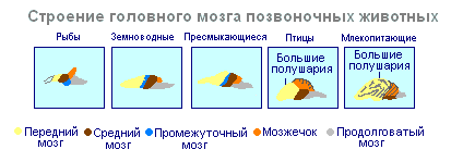
In all classes of vertebrate animals, the brain consists of 5 sections (anterior, intermediate, middle, posterior and medulla), but the degree of their development is not the same in animals of different classes.
Thus, in cyclostomes, all parts of the brain are located one after another in a horizontal plane. The medulla oblongata directly passes into the spinal cord with the central canal in the nutria.
In fish, the brain is more differentiated compared to cyclostomes. The volume of the forebrain is increased, especially in lungfishes, but the forebrain is not yet divided into hemispheres and functionally serves as the highest olfactory center. The roof of the forebrain is thin, it consists only of epithelial cells and does not contain nervous tissue. In the diencephalon, with which the pineal and pituitary glands are connected, the hypothalamus is located, which is the center of the endocrine system. The most developed in fish is the midbrain. The optic lobes are well expressed in it. In the region of the midbrain there is a bend characteristic of all higher vertebrates. In addition, the midbrain is an analyzing center. The cerebellum, which is part of the hindbrain, is well developed due to the complexity of movement in fish. It represents the center of coordination of movement, its size varies depending on the activity of movement of different species of fish. The medulla oblongata provides communication between the higher parts of the brain and the spinal cord and contains the centers of respiration and circulation.
10 pairs of cranial nerves emerge from the fish brain.
This type of brain, in which the highest center of integration is the midbrain, is called ichthyopsid.
In amphibians, the nervous system in its structure is close to the nervous system of lungfishes, but is distinguished by significant development and complete separation of paired elongated hemispheres, as well as weak development of the cerebellum, which is due to the low mobility of amphibians and the monotony of their movements. But amphibians developed a roof for the forebrain, called the primary medullary vault - archipallium. The number of cranial nerves, like in fish, is ten. And the type of brain is the same, i.e. ichthyopsid.
Thus, all anamnia (cyclostomes, fish and amphibians) have an ichthyopsid type of brain.
In the structure of the brain of reptiles belonging to higher vertebrates, i.e. to amniotes, the features of a progressive organization are clearly expressed. The forebrain hemispheres have a significant predominance over other parts of the brain. At their base there are large accumulations of nerve cells - striatum. Islands of the old cortex, the archicortex, appear on the lateral and medial sides of each hemisphere. The size of the midbrain is reduced, and it loses its importance as a leading center. The bottom of the forebrain becomes the analyzing center, i.e. striped bodies. This type of brain is called sauropsid or striatal. The cerebellum is increased in size due to the variety of movements of reptiles. The medulla oblongata forms a sharp bend, characteristic of all amniotes. There are 12 pairs of cranial nerves leaving the brain.
The same type of brain is characteristic of birds, but with some features. The forebrain hemispheres are relatively large. The olfactory lobes in birds are poorly developed, which indicates the role of smell in the life of birds. In contrast, the midbrain is represented by large optic lobes. The cerebellum is well developed, 12 pairs of nerves emerge from the brain.
The brain in mammals reaches its maximum development. The hemispheres are so large that they cover the midbrain and cerebellum. The cerebral cortex is especially developed, its area is increased due to convolutions and grooves. The cortex has a very complex structure and is called the new cortex - neocortex. A secondary medullary vault, the neopallium, appears. Large olfactory lobes are located in front of the hemispheres. The diencephalon, like other classes, includes the pineal gland, pituitary gland and hypothalamus. The midbrain is relatively small, it consists of four tubercles - the quadrigeminal. The anterior cortex is connected with the visual analyzer, the posterior one with the auditory one. Along with the forebrain, the cerebellum progresses greatly. There are 12 pairs of cranial nerves leaving the brain. The analyzing center is the cerebral cortex. This type of brain is called mammary.
3.4. Anomalies and malformations of the nervous system in humans.
1. Acephaly- absence of the brain, vault, skull and facial skeleton; this disorder is associated with underdevelopment of the anterior neural tube and is combined with defects of the spinal cord, bones and internal organs.
2. Anencephaly- absence of the cerebral hemispheres and skull roof with underdevelopment of the brain stem and is combined with other developmental defects. This pathology is caused by non-closure (dysraphism) of the head of the neural tube. In this case, the bones of the roof of the skull do not develop, and the bones of the base of the skull show various anomalies. Anencephaly is incompatible with life, the average frequency is 1/1500, and is more common in female fetuses.
3. Atelencephaly– arrest of development (heterochrony) of the anterior part of the neural tube at the stage of three vesicles. As a result, the cerebral hemispheres and subcortical nuclei are not formed.
4. Prosencephaly– the telencephalon is divided by a longitudinal groove, but in depth both hemispheres remain connected to each other.
5. Holoprosencephaly– the telencephalon is not divided into hemispheres and has the appearance of a hemisphere with a single cavity (ventricle).
6. Alobar prosencephaly– division of the telencephalon is only in the posterior part, and the frontal lobes remain undivided.
7. Aplasia or hypoplasia of the corpus callosum– complete or partial absence of a complex commissure of the brain, i.e. corpus callosum.
8. Hydroencephaly- atrophy of the cerebral hemispheres in combination with hydrocephalus.
9. Agiriya- complete absence of grooves and convolutions (smooth brain) of the cerebral hemispheres.
10. Microgyria- reduction in the number and volume of furrows.
11. Congenital hydrocephalus- obstruction of part of the ventricular system of the brain and its outputs, it is caused by a primary disorder of the development of the nervous system.
12. Spina bifida- a defect in the closure and separation of the spinal neural tube from the skin ectoderm. Sometimes this anomaly is accompanied by diplomyelia, in which the spinal cord is split along a certain length into two parts, each with its own central recess.
13. Iniencephaly- a rare anomaly, incompatible with life, occurs more often in female fetuses. This is a gross anomaly of the back of the head and brain. The head is turned so that the face is facing upward. Dorsally, the scalp continues into the skin of the lumbodorsal or sacral region.
 The meaning and origin of the surname Belyaev
 Meshcheryakova's technique "English for children"
 The Snow Queen The Snow Queen audio