Functions and types of nerve fibers. General plan of the structure of a synapse
Conduction of nerve impulses along nerve fibers and through synapses. The high-voltage potential that occurs when a receptor is excited in a nerve fiber is 5-10 times greater than the threshold for receptor stimulation. The conduction of the excitation wave along the nerve fiber is ensured by the fact that each subsequent section is irritated by the high-voltage potential of the previous section. In the pulpy nerve fibers, this potential does not spread continuously, but spasmodically; it jumps over one or even several interceptions of Ranvier, in which it intensifies. The duration of excitation between two adjacent nodes of Ranvier is equal to 5-10% of the duration of the high-voltage potential.
Carrying out nerve impulse along the nerve fiber occurs only under the condition of its anatomical continuity and its normal physiological state. Violation physiological properties nerve fiber, severe cooling or poisoning with poisons and drugs stops the conduction of the nerve impulse even with its anatomical continuity.
Nerve impulses are carried out in isolation along separate motor and sensory nerve fibers that are part of the mixed nerve, which depends on the insulating properties of the myelin sheaths covering them. In non-pulp nerve fibers, the biocurrent spreads continuously along the fiber and, thanks to the connective tissue sheath, does not pass from one fiber to another. A nerve impulse can travel along a nerve fiber in two directions: centripetal and centrifugal. Consequently, there are three rules for conducting a nerve impulse in nerve fibers: 1) anatomical continuity and physiological integrity, 2) isolated conduction and 3) bilateral conduction.
2-3 days after separation nerve fibers from the body of the neuron, they begin to degenerate, or degenerate, and the conduction of nerve impulses stops. Nerve fibers and myelin are destroyed and only the connective tissue sheath is preserved. If you connect the cut ends of the nerve fibers, or nerve, then after degeneration of those areas that are separated from nerve cells, restoration, or regeneration, of nerve fibers begins from the neuron bodies, from which they grow into the preserved connective tissue membranes. Regeneration of nerve fibers leads to restoration of impulse conduction.
Unlike nerve fibers through neurons nervous system nerve impulses are conducted in only one direction - from the receptor to the working organ. This depends on the nature of the nerve impulse through the synapses. In the nerve fiber above the presynaptic membrane there are many tiny acetylcholine vesicles. When the biocurrent reaches the presynaptic membrane, some of these vesicles burst, and acetylcholine passes through the smallest holes in the presynaptic membrane into the synaptic cleft.
There are areas in the postsynaptic membrane that have a special affinity for acetylcholine, which causes the temporary appearance of pores in the postsynaptic membrane, causing it to become temporarily permeable to ions. As a result, excitation and a high-voltage potential arise in the postsynaptic membrane, which spreads through the next neuron or through the innervated organ. Consequently, the transmission of excitation through synapses occurs chemically through an intermediary, or transmitter, acetylcholine, and the conduction of excitation to the next neuron is again carried out electrically.
The effect of acetylcholine on the conduction of nerve impulses through the synapse is short-lived; it is quickly destroyed and hydrolyzed by the enzyme cholinesterase.
Since the chemical transmission of a nerve impulse at a synapse occurs within a fraction of a millisecond, the nerve impulse at each synapse is delayed for this time.
Unlike nerve fibers, in which information is transmitted according to the “all or nothing” principle, i.e. discretely, in synapses information is transmitted according to the “more or less” principle, i.e. gradually. The more acetylcholine mediator is formed to a certain limit, the higher the frequency of high-voltage potentials in the subsequent neuron. After this limit, excitation turns into inhibition. Thus, digital information transmitted along nerve fibers is transformed into measurement information at synapses. Electronic measuring machines,
in which there are certain relationships between actually measured quantities and the quantities that they represent, are called analog, working on the principle of “more or less”; we can assume that a similar process occurs in synapses and its transition to digital occurs. Therefore, the nervous system functions according to mixed type: both digital and analog processes take place in it.
Action potential or nerve impulse specific reaction, flowing in the form of an exciting wave and flowing throughout neural pathway. This reaction is a response to a stimulus. The main task is the transmission of data from the receptor to the nervous system, and after that it sends this information to the desired muscles, glands and tissues. After the passage of the pulse, the surface part of the membrane becomes negatively charged, while its inner part remains positive. Thus, a nerve impulse is a sequentially transmitted electrical change.
The stimulating effect and its spread is subject to physico-chemical nature. The energy for this process is generated directly in the nerve itself. This happens due to the fact that the passage of an impulse leads to the formation of heat. Once it has passed, the attenuation or reference state begins. In which only a fraction of a second the nerve cannot conduct a stimulus. The speed at which the pulse can be delivered ranges from 3 m/s to 120 m/s.
The fibers through which excitation passes have a specific sheath. Roughly speaking, this system resembles electrical cable. The composition of the membrane can be myelin or non-myelin. The most important component of the myelin sheath is myelin, which plays the role of a dielectric.
The speed of the pulse depends on several factors, for example, on the thickness of the fibers; the thicker it is, the faster the speed develops. Another factor in increasing conduction speed is the myelin itself. But at the same time, it is not located over the entire surface, but in sections, as if strung together. Accordingly, between these areas there are those that remain “bare”. They cause current leakage from the axon.
An axon is a process that is used to transmit data from one cell to the rest. This process is regulated by a synapse - a direct connection between neurons or a neuron and a cell. There is also a so-called synaptic space or cleft. When an irritating impulse arrives at a neuron, neurotransmitters (molecules of a chemical composition) are released during the reaction. They pass through the synaptic opening, eventually reaching the receptors of the neuron or cell to which the data needs to be conveyed. Calcium ions are necessary for the conduction of a nerve impulse, since without this the neurotransmitter cannot be released.
The autonomic system is provided mainly by non-myelinated tissues. Excitement spreads through them constantly and continuously.
The transmission principle is based on the occurrence electric field, therefore, a potential arises that irritates the membrane of the adjacent section and so on throughout the fiber.
In this case, the action potential does not move, but appears and disappears in one place. The transmission speed through such fibers is 1-2 m/s. 
Laws of conduct
There are four basic laws in medicine:
- Anatomical and physiological value. Excitation is carried out only if there is no violation in the integrity of the fiber itself. If unity is not ensured, for example, due to infringement, drug use, then the conduction of a nerve impulse is impossible.
- Isolated conduction of irritation. Excitation can be transmitted along, in no way, without spreading to neighboring ones.
- Bilateral conduction. The path of impulse conduction can be of only two types - centrifugal and centripetal. But in reality, the direction occurs in one of the options.
- Non-decremental implementation. The impulses do not subside, in other words, they are carried out without decrement.
Chemistry of impulse conduction
The irritation process is also controlled by ions, mainly potassium, sodium and some organic compounds. The concentration of these substances is different, the cell is negatively charged inside itself, and positively charged on the surface. This process will be called potential difference. When hesitating negative charge, for example, its decrease provokes a potential difference and this process is called depolarization.

Stimulation of a neuron entails the opening of sodium channels at the site of stimulation. This may facilitate the entry of positively charged particles into the cell. Accordingly, the negative charge is reduced and an action potential or nerve impulse occurs. After this, the sodium channels close again.
It is often found that it is the weakening of polarization that promotes the opening of potassium channels, which provokes the release of positively charged potassium ions. This action reduces the negative charge on the cell surface.
The resting potential or electrochemical state is restored when potassium-sodium pumps are activated, with the help of which sodium ions leave the cell and potassium ions enter it.
As a result, we can say that when electrochemical processes are resumed, impulses occur that travel along the fibers.
319. Conduction of nerve impulses
A nerve impulse is an excitation wave propagating along a nerve fiber that occurs when a neuron is stimulated and carries a signal about a change in the environment (centripetal impulse) or a command signal in response to a change that has occurred (centrifugal impulse).
Resting potential. The occurrence and conduction of an impulse is associated with a change in the state of some structural elements of the neuron. These structures include a sodium pump, including Na^1^-ATPase, and two types of ion-conducting channels - sodium and potassium. Their interaction gives, in a state of rest, a potential difference across different sides plasma membrane of axons (resting potential). The existence of a potential difference is due to 1) the high concentration of potassium ions in the cell (20-50 times higher than in the environment); 2) the fact that intracellular anions (proteins and nucleic acids) cannot leave the cell; 3) with the fact that the permeability of the membrane for sodium ions is 20 times lower than for potassium ions. The potential exists ultimately because potassium ions tend to leave the cell to equalize external and internal concentrations. But potassium ions cannot leave the cell, and this leads to the appearance of a negative charge, which inhibits further equalization of the concentrations of potassium ions.
Chlorine ions must remain outside to compensate for the charge of poorly penetrating sodium, but tend to leave the cell along the concentration gradient. For supporting membrane potential (about 75 mV), it is necessary to maintain the difference in the concentrations of sodium and potassium ions so that sodium ions entering the cell are removed back from it in exchange for potassium ions. "This is achieved due to the action of membrane Na +, g^-ATPase, which, due to the energy of ATP, transfers sodium ions from the cell in exchange for two potassium ions taken into the cell. When abnormal high concentration sodium ions in external environment
the pump increases the Na + /K + ratio. Thus, at rest, potassium ions move outward along the gradient. At the same time, a certain amount of potassium is returned by diffusion. The difference between these processes is compensated by the action of the K" 1 ", N8"" pump. Sodium ions enter along a gradient at a rate limited by the permeability of the membrane for them. At the same time, sodium ions are pumped out by a pump against the concentration gradient due to the energy of ATP. Action potential - sequence of processes caused in a nerve by a stimulus. Irritation of the nerve entails local depolarization of the membrane, a decrease in membrane potential. This occurs due to the entry of a certain amount of sodium ions into the cell. When the potential difference drops to threshold level
The duration of the action potential is less than 1 ms and covers (unlike the resting potential) only a small portion of the axon.
In myelinated fibers, this is the area between adjacent nodes of Ranve. If the resting potential has changed to a degree that does not reach the threshold, then the action potential does not arise, but if the threshold value is reached, then in each case the same action potential develops (again, “all or nothing”).
The movement of potential in unmyelinated axons occurs as follows.
The diffusion of ions from an area with inverted polarity to neighboring ones causes the development of an action potential in them. In this regard, having arisen in one place, the potential spreads along the entire length of the axon.
The movement of an action potential is a nerve impulse, or a propagating wave of excitation, or conduction. Changes in the concentration of calcium ions inside the axons may be associated with the movement of the action potential and its conduction. All intracellular calcium, except for a small fraction, is bound to protein (the concentration of free calcium is about 0.3 mM), while around the cell its concentration reaches 2 mM. Therefore, there is a gradient that tends to push calcium ions into the cell. The nature of the calcium extrusion pump is unclear.
It is known, however, that each calcium ion is exchanged for 3 sodium ions, which penetrate the cell at the moment the action potential increases.
Sodium channel structure
Spontaneous release of potassium from the cell occurs through independent channels, the diameter of which is about
The threshold level of membrane potential at which its permeability to sodium increases depends on the calcium concentration outside the cell; its decrease during hypocalcemia causes convulsions.
The occurrence of an action potential and the propagation of an impulse in an unmyelinated nerve occurs due to the opening of a sodium channel. The channel is formed by integral protein molecules, its conformation changes in response to an increase in positive charge environment. The increase in charge is associated with the entry of sodium through the adjacent channel.
Depolarization caused by channel opening effectively affects the adjacent channel
In the myelinated nerve, sodium channels are concentrated in the unmyelinated nodes of Ranvier (more than ten thousand per 1 micron). In this regard, in the interception zone, the sodium flow is 10-100 times greater than on the conducting surface of the unmyelinated nerve. Na^K^-ATPase molecules in large quantities are located on adjacent sections of the nerve.
Depolarization of one of the nodes causes a potential gradient between the nodes, so the current quickly flows through the axoplasm to the adjacent node, reducing the potential difference there to a threshold level. This ensures a high speed of impulse transmission along the nerve - no less than 2 times faster than through a non-myelinated nerve (up to 50 m/s in a non-myelinated nerve and up to 100 m/s in a myelinated one). , 320.Transmission of nerve impulses - those. its spread to another cell is carried out using special structures synapses
, connecting the nerve ending and the neighboring cell. The synaptic cleft separates the cells. If the gap width is below 2 nm, signal transmission occurs by current propagation, as along the axon. In most synapses, the gap width approaches 20 nm. In these synapses, the arrival of an action potential leads to the release of a transmitter substance from the presynaptic membrane, which diffuses through the synaptic gap and binds to a specific receptor on the postsynaptic membrane, transmitting a signal to it. Mediator substances (neurotransmitters) - compounds that are in the presynaptic structure in sufficient concentration are released when, cause an electrical impulse after binding to the postsynaptic membrane. An essential feature of a neurotransmitter is the presence of a transport system for its removal from the synapse. Moreover, this transport system must have a high affinity for the neurotransmitter.
Depending on the nature of the mediator providing synaptic transmission, synapses are distinguished as cholinergic (transmitter - acetylcholine), and adrenergic (transmitters - catecholamine, norepinephrine, dopamine and, possibly, adrenaline)
Neurotransmitters– these are substances that are characterized by the following characteristics:
Accumulate in the presynaptic membrane in sufficient concentration;
Released upon transmission of impulse;
After binding to the postsynaptic membrane, they cause a change in speed metabolic processes and the occurrence of an electrical impulse;
They have an inactivation system or a transport system for removing hydrolysis products from the synapse.
Neurotransmitters play important role in functioning nerve tissue, providing synaptic transmission of nerve impulses. Their synthesis occurs in the body of neurons, and accumulation in special vesicles, which gradually move with the participation of systems of neurofilaments and neurotubules to the tips of axons.
Neurotransmitters include amino acid derivatives: taurine, norepinephrine, dopamine, GABA, glycine, acetylcholine, homocysteine and some others (adrenaline, serotonin, histamine), as well as neuropeptides.
Cholinergic synapses
Acetylcholine synthesized from choline and acetyl-CoA. Choline synthesis requires the amino acids serine and methionine. But, as a rule, ready-made choline enters the nervous tissue from the blood. Acetylcholine is involved in synaptic transmission of nerve impulses. It accumulates in synaptic vesicles, forming complexes with the negatively charged protein vesiculin (Fig. 22). The transfer of excitation from one cell to another is carried out using a special synaptic mechanism.
Rice. 22. Cholinergic synapse
A synapse is a functional contact of specialized areas plasma membranes two excitable cells. A synapse consists of a presynaptic membrane, a synaptic cleft, and a postsynaptic membrane. The membranes at the point of contact have thickenings in the form of plaques - nerve endings. A nerve impulse that reaches a nerve ending is unable to overcome the obstacle that has arisen in front of it - the synaptic cleft. After this, the electrical signal is converted into a chemical one.
The presynaptic membrane contains special channel proteins similar to the proteins that form the sodium channel in the axon membrane. They also respond to the membrane potential by changing their conformation and forming a channel. As a result, Ca 2+ ions pass through the presynaptic membrane along a concentration gradient into the nerve ending. The Ca 2+ concentration gradient is created by the work of the Ca 2+ -dependent ATPase. An increase in the concentration of Ca 2+ inside the nerve ending causes the fusion of acetylcholine-filled vesicles present there. Acetylcholine is then secreted into the synaptic cleft by exocytosis and binds to receptor proteins located on the surface of the postsynaptic membrane.
The acetylcholine receptor is a transmembrane oligomeric glycoprotein complex consisting of 6 subunits. The density of receptor proteins in the postsynaptic membrane is very high - about 20,000 molecules per 1 µm 2. The spatial structure of the receptor strictly corresponds to the conformation of the mediator. When interacting with acetylcholine, the receptor protein changes its conformation so that a sodium channel is formed inside it. The cation selectivity of the channel is ensured by the fact that the gate of the channel is formed by negatively charged amino acids. That. The permeability of the postsynaptic membrane to sodium increases and an impulse (or contraction of the muscle fiber) occurs. Depolarization of the postsynaptic membrane causes dissociation of the acetylcholine-protein-receptor complex, and acetylcholine is released into the synaptic cleft. Once acetylcholine is in the synaptic cleft, it undergoes rapid hydrolysis within 40 μs by the enzyme acetylcholinesterase into choline and acetyl-CoA.
Irreversible inhibition of acetylcholinesterase causes death. Enzyme inhibitors are organophosphorus compounds. Death occurs as a result of respiratory arrest. Reversible acetylcholinesterase inhibitors are used as therapeutic drugs, for example, in the treatment of glaucoma and intestinal atony.
Adrenergic synapses(Fig. 23) are found in postganglionic fibers, in fibers of the sympathetic nervous system, in various parts of the brain. They serve as mediators catecholamines: norepinephrine and dopamine. Catecholamines in nervous tissue are synthesized according to a general mechanism from tyrosine. The key enzyme in the synthesis is tyrosine hydroxylase, which is inhibited by the end products.

Rice. 23. Adrenergic synapse
Norepinephrine– a mediator in postganglionic fibers of the sympathetic system and in various parts of the central nervous system.
Dopamine– a mediator of pathways, the bodies of neurons of which are located in the brain. Dopamine is responsible for controlling voluntary movements. Therefore, when dopaminergic transmission is disrupted, the disease parkinsonism occurs.
Catecholamines, like acetylcholine, accumulate in synaptic vesicles and are also released into the synaptic cleft upon receipt of a nerve impulse. But regulation in the adrenergic receptor occurs differently. The presynaptic membrane has a special regulatory protein– achromogranin, which, in response to an increase in the concentration of the transmitter in the synaptic cleft, binds the already released transmitter and stops its further exocytosis. There is no enzyme that destroys the transmitter in adrenergic synapses. After the impulse is transmitted, the mediator molecules are pumped by a special transport system by active transport with the participation of ATP back into the presynaptic membrane and are reincorporated into vesicles. In the presynaptic nerve ending excess mediator can be inactivated by monoamine oxidase (MAO), as well as catecholamine-O-methyltransferase (COMT) by methylation at the hydroxy group.
Signal transmission at adrenergic synapses occurs with the participation of the adenylate cyclase system. Binding of the transmitter to the postsynaptic receptor almost instantly causes an increase in the concentration of cAMP, which leads to rapid phosphorylation of proteins of the postsynaptic membrane. As a result, the generation of nerve impulses in the postsynaptic membrane is inhibited. In some cases, the immediate cause of this is an increase in the permeability of the postsynaptic membrane for potassium, or a decrease in conductivity for sodium (this condition leads to hyperpolarization).
Taurine formed from the amino acid cysteine. First, sulfur in the HS group is oxidized (the process occurs in several stages), then decarboxylation occurs. Taurine is an unusual acid in which there is no carboxyl group, but a sulfuric acid residue. Taurine takes part in the conduction of nerve impulses in the process of visual perception.
GABA – inhibitory transmitter (about 40% of neurons). GABA increases the permeability of postsynaptic membranes for potassium ions. This leads to a change in membrane potential. GABA inhibits the inhibition of “unnecessary” information: attention, motor control.
Glycine– auxiliary inhibitory transmitter (less than 1% of neurons). In terms of the effects it causes, it is similar to GABA. Its function is inhibition of motor neurons.
Glutamic acid- the main excitatory transmitter (about 40% of neurons). Main function: conducting the main flows of information in the central nervous system (sensory signals, motor commands, memory).
Normal activity of the central nervous system is ensured by a delicate balance of glutamic acid and GABA. Violation of this balance (usually in the direction of decreasing inhibition) negatively affects many nervous processes. When the balance is disturbed, attention deficit hyperactivity disorder in children (ADHD) develops, nervousness and anxiety in adults, sleep disturbance, insomnia, and epilepsy increase.
Neuropeptides contain from three to several dozen amino acid residues. They function only in the higher parts of the nervous system. These peptides function not only as neurotransmitters, but also as hormones. They transmit information from cell to cell through the circulation system. These include:
Neurohypophyseal hormones (vasopressin, liberins, statins) - they are both hormones and mediators;
Gastrointestinal peptides (gastrin, cholecystokinin). Gastrin causes a feeling of hunger, cholecystokinin causes a feeling of fullness, and also stimulates contraction of the gallbladder and pancreatic function;
Opiate-like peptides (or analgesic peptides). They are formed through reactions of limited proteolysis of the proopiocortin precursor protein. Interacts with the same receptors as opiates (for example, morphine), thereby imitating their action. Common name- endorphins. They are easily destroyed by proteinases, so their pharmacological effect is negligible;
Sleep peptides. Their molecular nature has not been established. They induce sleep;
Memory peptides (scotophobin). Accumulates during training to avoid darkness;
Peptides are components of the renin-angiotensin system. Stimulate the thirst center and the secretion of antidiuretic hormone.
The formation of peptides occurs as a result of limited proteolysis reactions; they are destroyed under the action of proteinases.
1. Describe chemical composition brain.
2. What are the features of metabolism in nervous tissue?
3. List the functions of glutamate in nervous tissue.
4. What is the role of mediators in the transmission of nerve impulses? List the main inhibitory and excitatory mediators.
5. What are the differences in the functioning of adrenergic and cholinergic synapses?
6. Give examples of compounds that affect the synaptic transmission of nerve impulses.
7. What biochemical changes can be observed in nervous tissue during mental illness?
8. What are the features of the action of neuropeptides?
Biochemistry of muscle tissue
Muscles make up 40-50% of a person’s body weight.
Distinguish three types of muscles:
Striated skeletal muscles (contract voluntarily);
Striated cardiac muscle (contracts involuntarily);
Smooth muscles (vessels, intestines, uterus) (contract involuntarily).
Striated muscle consists of numerous elongated fibers.
Muscle fiber- multinucleate cell covered with an elastic membrane - sarcolemma. Muscle fiber contains motor nerves, transmitting to it a nerve impulse that causes contraction. Along the length of the fiber in semi-liquid sarcoplasm thread-like formations are located - myofibrils. Sarcomere- repeating element of myofibril, limited by Z-line(Fig. 24). In the middle of the sarcomere there is an A-disc, dark in a phase-contrast microscope, in the center of which there is an M-line, visible under electron microscopy. The H-zone occupies the middle part
A-disc. I-disks are bright under a phase-contrast microscope, and each of them is divided into equal halves by a Z-line. A-discs contain thick myosin and thin actin filaments. Thin filaments begin at the Z-line, pass through the I-disc and are interrupted in the H-zone. Electron microscopy showed that thick filaments are arranged in a hexagonal shape and extend across the entire A-disc. Between the thick threads are thin ones. When the muscle contracts, the I-discs practically disappear, and the area of overlap between the thin and thick filaments increases.
Sarcoplasmic reticulum- an intracellular membrane system of interconnected flattened vesicles and tubules that surrounds the sarcomeres of myofibrils. Its inner membrane contains proteins that can bind calcium ions.
Nerve fiber structure. The conduction of nerve impulses is a specialized function of nerve fibers, i.e. processes of nerve cells.
Nerve fibers separate soft, or myelinated, And pulpless, or unmyelinated. Pulp, sensory and motor fibers are part of the nerves supplying the sensory organs and skeletal muscles; they are also present in the autonomic nervous system. Non-pulp fibers in vertebrates belong mainly to the sympathetic nervous system.
Nerves usually consist of both pulpy and non-pulphate fibers, and their ratio in different nerves is different. For example, in many cutaneous nerves the predominant nerve fibers predominate. Thus, in the nerves of the autonomic nervous system, for example in the vagus nerve, the number of soft fibers reaches 80-95%. In contrast, the nerves innervating skeletal muscles contain only a relatively small number of non-pulp fibers.
As electron microscopic studies have shown, the myelin sheath is created as a result of the fact that the myelocyte (Schwann cell) repeatedly wraps the axial cylinder (Fig. 2.27"), its layers merge, forming a dense fatty sheath - the myelin sheath. The myelin sheath through gaps equal length is interrupted, leaving open areas of the membrane approximately 1 µm wide. These areas were named Ranvier interceptions.
Rice. 2.27. The role of the myelocyte (Schwann cell) in the formation of the myelin sheath in the pulpy nerve fibers: successive stages of the spiral-shaped twisting of the myelocyte around the axon (I); mutual arrangement myelocytes and axons in non-pulp nerve fibers (II)
The length of the interstitial areas covered by the myelin sheath is approximately proportional to the diameter of the fiber. Thus, in nerve fibers with a diameter of 10-20 microns, the length of the gap between the interceptions is 1-2 mm. In the thinnest fibers (diameter
1-2 µm) these areas are about 0.2 mm long.
Non-pulp nerve fibers do not have a myelin sheath; they are isolated from each other only by Schwann cells. In the simplest case, a single myelocyte surrounds one pulpless fiber. Often, however, several thin, pulpless fibers appear in the folds of the myelocyte.
The myelin sheath has a dual function: an electrical insulator function and a trophic function. The insulating properties of the myelin sheath are due to the fact that myelin, as a substance of lipid nature, prevents the passage of ions and therefore has a very high resistance. Due to the existence of the myelin sheath, the occurrence of excitation in the pulpal nerve fibers is not possible throughout the entire length of the axial cylinder, but only in limited areas - the nodes of Ranvier. This is important for the propagation of the nerve impulse along the fiber.
The trophic function of the myelin sheath, apparently, is that it takes part in the processes of regulation of metabolism and growth of the axial cylinder.
Conduction of excitation in unmyelinated and myelinated nerve fibers. In soft nerve fibers, excitation spreads continuously along the entire membrane, from one excited area to another located nearby. In contrast, in myelinated fibers the action potential can propagate only spasmodically, “jumping” through sections of the fiber covered with an insulating myelin sheath. This is called salipatory.
Direct electrophysiological studies carried out by Kato (1924) and then by Tasaki (1953) on single myelinated frog nerve fibers showed that action potentials in these fibers arise only at the nodes, and the myelin-covered areas between the nodes are practically inexcitable.
The density of sodium channels in the interceptions is very high: there are about 10,000 sodium channels per 1 μm2 membrane, which is 200 times higher than their density in the membrane of the giant squid axon. High density sodium channels are the most important condition saltatory conduction of excitation. In Fig. Figure 2.28 shows how a nerve impulse “jumps” from one interception to another.
At rest, the outer surface of the excitable membrane of all nodes of Ranvier is positively charged. There is no potential difference between adjacent interceptions. At the moment of excitation, the surface of the interception membrane WITH becomes charged electronegatively with respect to the membrane surface of the adjacent interception D. This leads to the emergence of local (lo 
Rice. 2.28.
A- Not myelinated fiber; IN- myelinated fiber. The arrows show the direction of the current
cal) electric current, which goes through the interstitial fluid surrounding the fiber, membrane and axoplasm in the direction shown in the figure by the arrow. Coming out through interception D the current excites it, causing the membrane to recharge. In interception WITH the excitement still continues, and he becomes refractory for a while. Therefore interception D is capable of bringing into a state of excitation only the next interception, etc.
“Jumping” of the action potential across the interinterceptor region is possible only because the amplitude of the action potential in each interception is 5-6 times higher than the threshold value required to excite the neighboring interception. Under certain conditions, the action potential can “jump” not only through one, but also through two interinterceptor sections - in particular, if the excitability of the adjacent interception is reduced by some pharmacological agent, for example, novocaine, cocaine, etc.
The assumption about the spasmodic propagation of excitation in nerve fibers was first expressed by B.F. Verigo (1899). This method of conduction has a number of advantages compared to continuous conduction in non-pulp fibers: firstly, by “jumping” over relatively large sections of the fiber, excitation can spread at a much higher speed than with continuous conduction along a non-pulp fiber of the same diameter; secondly, abrupt propagation is energetically more economical, since not the entire membrane comes into a state of activity, but only its small sections in the interception area, having a width of less than 1 μm. The losses of ions (per unit length of fiber) accompanying the occurrence of an action potential in such limited areas of the membrane are very small, and therefore the energy costs for work are also low. sodium-potassium pump, necessary to restore altered ionic ratios between the internal contents of the nerve fiber and tissue fluid.
- See: Human Physiology / Ed. A. Kositsky.
 Mikhail Lozinsky where he published
 Functional linguistics The concept of functional linguistics
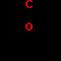 Preparation and use of aldehydes and ketones