What nerve is there in the human body. What is the nervous system? The activity of the nervous system, condition and protection
The nervous system is the highest integrating and coordinating system of the human body, which ensures the coordinated activity of internal organs and the connection of the body with the external environment.
Anatomically, the nervous system is divided into the central (brain and spinal cord); and peripheral, including 12 pairs of cranial nerves, 31 pairs of spinal nerves and nerve nodes located outside the brain and spinal cord.
The function of the nervous system is divided into:
the somatic nervous system - mainly carries out the connection of the body with the external environment: the perception of irritations, the regulation of movements of the striated muscles, etc.
autonomic (autonomous) nervous system - regulates metabolism and the functioning of internal organs: heartbeat, vascular tone, peristaltic contractions of the intestine, secretion of various glands, etc. The autonomic nervous system is divided into parasympathetic and sympathetic nervous systems.
Both of them function in close interaction, however, the autonomic nervous system has some independence, controlling involuntary functions.
The nervous system is made up of nerve cells called neurons. There are 25 billion neurons in the brain, and 25 million cells in the periphery. The bodies of neurons are located mainly in the CNS. Gray matter is a collection of neurons. In the spinal cord, it is located in the center, surrounding the spinal canal. In the brain, on the contrary, the gray matter is located on the surface, forming a cortex and separate clusters - nuclei concentrated in the white matter.
White matter is under gray and is composed of nerve fibers (neuronal processes) covered with sheaths. Nerve ganglions also consist of bodies of neurons. Nerve fibers that extend beyond the CNS and nerve nodes, connecting, compose nerve bundles, and several such bundles form individual nerves.
Centripetal, or sensitive - nerves that conduct excitation from the periphery to the central nervous system. For example, visual, olfactory, auditory.
Centrifugal, or motor - nerves through which excitation is transmitted from the central nervous system to the organs. For example, oculomotor.
Mixed (wandering, spinal), if excitation goes in one direction along one fiber, and in the other direction along the other.
Functions nervous system: regulates the activity of all organs and organ systems, communicates with the external environment through the sense organs; is the material basis for higher nervous activity, thinking, behavior and speech.
The structure and function of the spinal cord.
The spinal cord is located in the spinal canal from the 1st cervical vertebra to the 1st - 2nd lumbar, its length is about 45 cm, thickness is about 1 cm. The anterior and posterior longitudinal grooves divide it into two symmetrical halves. In the center is the spinal canal, which contains the cerebrospinal fluid. In the middle part of the spinal cord, near the spinal canal, there is gray matter, which in cross section resembles the contour of a butterfly. The gray matter is formed by the bodies of neurons, it distinguishes between the anterior and posterior horns. The bodies of intercalary neurons are located in the posterior horns of the spinal cord, and the bodies of motor neurons are located in the anterior horns. In the thoracic region, lateral horns are also distinguished, in which the neurons of the sympathetic part of the autonomic nervous system are located. Surrounding the gray matter is the white matter formed by the nerve fibers. The spinal cord is covered by three membranes:
hard shell - outer, connective tissue, lining the inner cavity of the skull and spinal canal;
arachnoid - located under the solid. This is a thin shell with a small number of nerves and vessels;
the choroid is fused with the brain, enters the furrows and contains many blood vessels.
Fluid-filled cavities form between the vascular and arachnoid membranes.
31 pairs of mixed spinal nerves leave the spinal cord. Each nerve begins with two roots: the anterior (motor), which contains the processes of motor neurons and autonomic fibers, and the posterior (sensory), through which excitation is transmitted to the spinal cord. In the posterior roots are the spinal nodes - clusters of sensory neuron bodies.
Transection of the posterior roots leads to a loss of sensation in those areas that are innervated by the corresponding roots, and transection of the anterior roots leads to paralysis of the innervated muscles.
The functions of the spinal cord are reflex and conduction. As a reflex center, the spinal cord takes part in motor (conducts nerve impulses to the skeletal muscles) and autonomic reflexes. The most important vegetative reflexes of the spinal cord are vasomotor, food, respiratory, defecation, urination, sexual. The reflex function of the spinal cord is under the control of the brain.
The reflex functions of the spinal cord can be examined on the spinal preparation of a frog (without a brain), which retains the simplest motor reflexes. She withdraws her paw in response to mechanical and chemical stimuli. In humans, the brain is of decisive importance in the implementation of the coordination of motor reflexes.
The conduction function is carried out due to the ascending and descending paths of the white matter. Excitation from the muscles and internal organs is transmitted along the ascending paths to the brain, along the descending paths - from the brain to the organs.
The structure and functions of the brain.
There are five sections in the brain: the medulla oblongata; the hindbrain, which includes the bridge and the cerebellum; midbrain; diencephalon and forebrain, represented by the large hemispheres. Up to 80% of the mass of the brain falls on the cerebral hemispheres. The central canal of the spinal cord continues into the brain, where it forms four cavities (ventricles). Two ventricles are located in the hemispheres, the third - in the diencephalon, the fourth - at the level of the medulla oblongata and the bridge. They contain cranial fluid. The brain, as well as the spinal cord, is surrounded by three membranes - connective tissue, arachnoid and vascular.
The medulla oblongata is a continuation of the spinal cord, performs reflex and conduction functions. Reflex functions are associated with the regulation of the work of the respiratory, digestive, and circulatory organs. Here are the centers of protective reflexes - coughing, sneezing, vomiting.
The bridge connects the cerebral cortex with the spinal cord and cerebellum, performing mainly a conductive function.
The cerebellum is formed by two hemispheres, externally covered with a bark of gray matter, under which is white matter. The white matter contains nuclei. The middle part of the cerebellum - the worm - connects its hemispheres. The cerebellum is responsible for coordination, balance and influences muscle tone. When the cerebellum is damaged, there is a decrease in muscle tone and a disorder in the coordination of movements, but after a while other parts of the nervous system begin to perform the functions of the cerebellum, and the lost functions are partially restored. Together with the bridge, the cerebellum is part of the hindbrain.
The midbrain connects all parts of the brain. Here are the centers of skeletal muscle tone, the primary centers of visual and auditory orienting reflexes, which are manifested in the movements of the eyes and head towards stimuli.
Three parts are distinguished in the diencephalon: the visual tubercles (thalamus), the epithalamic region (epithalamus), which includes the pineal gland, and the hypothalamic region (hypothalamus). The subcortical centers of all types of sensitivity are located in the thalamus, excitation from the sense organs comes here, and from here it is transmitted to various parts of the cerebral cortex. The hypothalamus contains the highest regulatory centers of the autonomic nervous system. It controls the constancy of the internal environment of the body. Here are the centers of appetite, thirst, sleep, thermoregulation, i.e. regulation of all types of metabolism. Neurons of the hypothalamus produce neurohormones that regulate the functioning of the endocrine system. In the diencephalon there are also emotional centers: centers of pleasure, fear, aggression. Together with the hindbrain and medulla, the diencephalon is part of the brainstem.
The forebrain is represented by the cerebral hemispheres connected by the corpus callosum. The surface of the forebrain is formed by the cortex, the area of which is about 2200 cm 2. Numerous folds, convolutions and furrows significantly increase the surface of the cortex. The surface of the convolutions is more than two times smaller than the surface of the furrows. The human cortex has from 14 to 17 billion nerve cells arranged in 6 layers, the thickness of the cortex is 2-4 mm. Accumulations of neurons in the depths of the hemispheres form subcortical nuclei. The cerebral cortex consists of 4 lobes: frontal, parietal, temporal and occipital, separated by furrows. In the cortex of each hemisphere, the central sulcus separates the frontal lobe from the parietal, the lateral sulcus separates the temporal lobe, and the parietal-occipital sulcus separates the occipital lobe from the parietal.
In the cortex, sensory, motor and associative zones are distinguished. Sensitive zones are responsible for the analysis of information coming from the sense organs: occipital - for vision, temporal - for hearing, smell and taste; parietal - for skin and joint-muscular sensitivity. Moreover, each hemisphere receives impulses from the opposite side of the body. The motor zones are located in the posterior regions of the frontal lobes, from here come the commands for contraction of the skeletal muscles, their defeat leads to muscle paralysis. Associative zones are located in the frontal lobes of the brain and are responsible for the development of programs for behavior and management of human labor activity; their mass in humans is more than 50% of the total mass of the brain.
A person is characterized by a functional asymmetry of the hemispheres: the left hemisphere is responsible for abstract-logical thinking, speech centers are also located there (Brock's center is responsible for pronunciation, Wernicke's center for understanding speech), the right hemisphere is for figurative thinking, musical and artistic creativity.
Due to the strong development of the cerebral hemispheres, the average mass of the human brain is 1400 g on average.
Include organs of the central nervous system (brain and spinal cord) and organs of the peripheral nervous system (peripheral ganglions, peripheral nerves, receptor and effector nerve endings).
Functionally, the nervous system is divided into somatic, which innervates skeletal muscle tissue, i.e., controlled by consciousness, and vegetative (autonomous), which regulates the activity of internal organs, blood vessels and glands, i.e. does not depend on consciousness.
The functions of the nervous system are regulatory and integrating.
It is laid on the 3rd week of embryogenesis in the form of a neural plate, which is transformed into a neural groove, from which a neural tube is formed. There are 3 layers in its wall:
Internal - ependymal:
Medium - raincoat. Later it turns into gray matter.
External - edge. It produces white matter.
In the cranial part of the neural tube, an extension is formed, from which 3 cerebral vesicles are formed at the beginning, and later - five. The latter give rise to five parts of the brain.
The spinal cord is formed from the trunk of the neural tube.
In the first half of embryogenesis, there is an intensive proliferation of young glial and nerve cells. Subsequently, a radial glia is formed in the mantle layer of the cranial region. Its thin long processes penetrate the wall of the neural tube. Young neurons migrate along these processes. There is a formation of centers of the brain (especially intensively from 15 to 20 weeks - a critical period). Gradually, in the second half of embryogenesis, proliferation and migration fade. After birth, division stops. When the neural tube is formed, cells that are located between the ectoderm and the neural tube are evicted from the neural folds (interlocking areas), forming the neural crest. The latter is split into 2 sheets:
1 - under the ectoderm, pigmentocytes (skin cells) are formed from it;
2 - around the neural tube - ganglionic plate. Peripheral nerve nodes (ganglia), the adrenal medulla, and sections of chromaffin tissue (along the spine) are formed from it. After birth, there is an intensive growth of the processes of nerve cells: axons and dendrites, synapses between neurons, neural circuits (a strictly ordered interneuronal connection) are formed, which make up reflex arcs (successively located cells that transmit information) that provide reflex activity of a person (especially the first 5 years of life child, so stimuli are needed to form bonds). Also in the first years of a child's life, myelination is the most intensive - the formation of nerve fibers.
PERIPHERAL NERVOUS SYSTEM (PNS).
Peripheral nerve trunks are part of the neurovascular bundle. They are mixed in function, contain sensory and motor nerve fibers (afferent and efferent). Myelinated nerve fibers predominate, and non-myelinated ones are in small quantities. Around each nerve fiber is a thin layer of loose connective tissue with blood and lymphatic vessels - endoneurium. Around the bundle of nerve fibers is a sheath of loose fibrous connective tissue - the perineurium - with a small number of vessels (it mainly performs a frame function). Around the entire peripheral nerve there is a sheath of loose connective tissue with larger vessels - the epineurium. Peripheral nerves regenerate well, even after complete damage. Regeneration is carried out due to the growth of peripheral nerve fibers. The growth rate is 1-2 mm per day (the ability to regenerate is a genetically fixed process).
spinal node
It is a continuation (part) of the posterior root of the spinal cord. Functionally sensitive. Outside covered with a connective tissue capsule. Inside - connective tissue layers with blood and lymphatic vessels, nerve fibers (vegetative). In the center - myelinated nerve fibers of pseudo-unipolar neurons located along the periphery of the spinal ganglion. Pseudo-unipolar neurons have a large rounded body, a large nucleus, well-developed organelles, especially the protein-synthesizing apparatus. A long cytoplasmic outgrowth departs from the body of the neuron - this is part of the body of the neuron, from which one dendrite and one axon depart. Dendrite - long, forms a nerve fiber that goes as part of a peripheral mixed nerve to the periphery. Sensitive nerve fibers end at the periphery with a receptor, i.e. sensitive nerve ending. Axons are short and form the posterior root of the spinal cord. In the posterior horns of the spinal cord, axons form synapses with interneurons. Sensitive (pseudo-unipolar) neurons constitute the first (afferent) link of the somatic reflex arc. All cell bodies are located in ganglia.
Spinal cord
Outside, it is covered with a pia mater, which contains blood vessels that penetrate into the substance of the brain. Conventionally, 2 halves are distinguished, which are separated by the anterior median fissure and the posterior median connective tissue septum. In the center is the central canal of the spinal cord, which is located in the gray matter, lined with ependyma, contains cerebrospinal fluid, which is in constant motion. Along the periphery is white matter, where there are bundles of nerve myelin fibers that form pathways. They are separated by glial-connective tissue septa. In the white matter, the anterior, lateral and posterior cords are distinguished.
In the middle part there is a gray matter, in which the posterior, lateral (in the thoracic and lumbar segments) and anterior horns are distinguished. The halves of the gray matter are connected by the anterior and posterior commissures of the gray matter. The gray matter contains a large number of glial and nerve cells. Gray matter neurons are divided into:
1) Internal neurons, completely (with processes) located within the gray matter, are intercalary and are located mainly in the posterior and lateral horns. There are:
a) Associative. located within one half.
b) Commissural. Their processes extend into the other half of the gray matter.
2) Beam neurons. They are located in the posterior horns and in the lateral horns. They form nuclei or are located diffusely. Their axons enter the white matter and form bundles of nerve fibers in an ascending direction. They are inserts.
3) Radicular neurons. They are located in the lateral nuclei (kernels of the lateral horns), in the anterior horns. Their axons extend beyond the spinal cord and form the anterior roots of the spinal cord.
In the superficial part of the posterior horns there is a spongy layer, which contains a large number of small intercalary neurons.
Deeper than this strip is a gelatinous substance containing mainly glial cells, small neurons (the latter in small quantities).
In the middle part is the own nucleus of the posterior horns. It contains large beam neurons. Their axons go to the white matter of the opposite half and form the dorsal-cerebellar anterior and dorsal-thalamic posterior pathways.
The cells of the nucleus provide exteroceptive sensitivity.
At the base of the posterior horns is the thoracic nucleus (Clark-Shutting column), which contains large bundle neurons. Their axons go to the white matter of the same half and participate in the formation of the posterior spinal cerebellar tract. Cells in this pathway provide proprioceptive sensitivity.
In the intermediate zone are the lateral and medial nuclei. The medial intermediate nucleus contains large bundle neurons. Their axons go to the white matter of the same half and form the anterior spinal cerebellar tract, which provides visceral sensitivity.
The lateral intermediate nucleus refers to the autonomic nervous system. In the thoracic and upper lumbar regions it is the sympathetic nucleus, and in the sacral region it is the nucleus of the parasympathetic nervous system. It contains an intercalary neuron, which is the first neuron of the efferent link of the reflex arc. This is a radicular neuron. Its axons exit as part of the anterior roots of the spinal cord.
In the anterior horns are large motor nuclei, which contain motor radicular neurons with short dendrites and a long axon. The axon exits as part of the anterior roots of the spinal cord, and then goes as part of the peripheral mixed nerve, represents motor nerve fibers and is pumped at the periphery by a neuromuscular synapse on skeletal muscle fibers. They are effectors. Forms the third effector link of the somatic reflex arc.
In the anterior horns, a medial group of nuclei is isolated. It is developed in the thoracic region and provides innervation to the muscles of the body. The lateral group of nuclei is located in the cervical and lumbar regions and innervates the upper and lower extremities.
In the gray matter of the spinal cord there is a large number of diffuse bundle neurons (in the posterior horns). Their axons go into the white matter and immediately divide into two branches that go up and down. Branches through 2-3 segments of the spinal cord return back to the gray matter and form synapses on the motor neurons of the anterior horns. These cells form their own apparatus of the spinal cord, which provides a connection between neighboring 4-5 segments of the spinal cord, which ensures the response of a muscle group (an evolutionarily developed protective reaction).
The white matter contains ascending (sensitive) pathways, which are located in the posterior cords and in the peripheral part of the lateral horns. Descending nerve pathways (motor) are located in the anterior cords and in the inner part of the lateral cords.
Regeneration. Very poorly regenerates gray matter. Regeneration of white matter is possible, but the process is very long.
Histophysiology of the cerebellum. The cerebellum refers to the structures of the brain stem, i.e. is a more ancient formation that is part of the brain.
Performs a number of functions:
balance;
The centers of the autonomic nervous system (ANS) (intestinal motility, blood pressure control) are concentrated here.
Outside covered with meninges. The surface is embossed due to deep furrows and convolutions, which are deeper than in the cerebral cortex (CBC).
On the cut is represented by the so-called "tree of life".
The gray matter is located mainly along the periphery and inside, forming nuclei.
In each gyrus, the central part is occupied by white matter, in which 3 layers are clearly visible:
1 - surface - molecular.
2 - medium - ganglionic.
3 - internal - granular.
1. The molecular layer is represented by small cells, among which basket and stellate (small and large) cells are distinguished.
Basket cells are located closer to the ganglion cells of the middle layer, i.e. inside the layer. They have small bodies, their dendrites branch in the molecular layer, in a plane transverse to the course of the gyrus. The neurites run parallel to the plane of the gyrus above the bodies of the pear-shaped cells (the ganglion layer), forming numerous branches and contacts with the dendrites of the pear-shaped cells. Their branches are braided around the bodies of pear-shaped cells in the form of baskets. Excitation of basket cells leads to inhibition of pear-shaped cells.
Outwardly, stellate cells are located, the dendrites of which branch out here, and the neurites participate in the formation of the basket and communicate by synapses with the dendrites and bodies of the pear-shaped cells.
Thus, the basket and stellate cells of this layer are associative (connecting) and inhibitory.
2. Ganglion layer. Here are located large ganglion cells (diameter = 30-60 microns) - Purkin' cells. These cells are located strictly in one row. The cell bodies are pear-shaped, there is a large nucleus, the cytoplasm contains EPS, mitochondria, the Golgi complex is poorly expressed. One neurite departs from the base of the cell, which passes through the granular layer, then into the white matter and ends at the cerebellar nuclei with synapses. This neurite is the first link in the efferent (descending) pathways. 2-3 dendrites depart from the apical part of the cell, which branch intensively in the molecular layer, while the branching of the dendrites occurs in a plane transverse to the course of the gyrus.
Pear-shaped cells are the main effector cells of the cerebellum, where an inhibitory impulse is produced.
3. Granular layer, saturated with cellular elements, among which cells - grains stand out. These are small cells, with a diameter of 10-12 microns. They have one neurite, which goes into the molecular layer, where it comes into contact with the cells of this layer. Dendrites (2-3) are short and branch into numerous "bird's foot" branches. These dendrites come into contact with afferent fibers called bryophytes. The latter also branch out and come into contact with the branching of the dendrites of cells - grains, forming glomeruli of thin weaves like moss. In this case, one mossy fiber is in contact with many cells - grains. And vice versa - the cell - the grain is also in contact with many mossy fibers.
Mossy fibers come here from the olives and the bridge, i.e. they bring here the information that comes through the associative neurons to the pear-shaped neurons. Large stellate cells are also found here, which lie closer to the pear-shaped cells. Their processes contact the granule cells proximal to the mossy glomeruli and in this case block the impulse transmission.
Other cells can also be found in this layer: stellate with a long neurite extending into the white matter and further into the adjacent gyrus (Golgi cells are large stellate cells).
Afferent climbing fibers - liana-like - enter the cerebellum. They come here as part of the spinal tracts. Then they crawl along the bodies of pear-shaped cells and along their processes, with which they form numerous synapses in the molecular layer. Here they carry an impulse directly to the pear-shaped cells.
Efferent fibers come out of the cerebellum, which are the axons of the piriform cells.
The cerebellum has a large number of glial elements: astrocytes, oligodendrogliocytes, which perform supporting, trophic, restrictive and other functions. A large amount of serotonin is released in the cerebellum, thus. the endocrine function of the cerebellum can also be distinguished.
Cerebral cortex (CBC)
This is a newer part of the brain. (It is believed that the CBP is not a vital organ.) It has great plasticity.
Thickness can be 3-5mm. The area occupied by the cortex increases due to furrows and convolutions. CBP differentiation ends by the age of 18, and then there are processes of accumulation and use of information. The mental abilities of an individual also depend on the genetic program, but in the end it all depends on the number of synaptic connections formed.
There are 6 layers in the cortex:
1. Molecular.
2. External granular.
3. Pyramidal.
4. Internal grainy.
5. Ganglionic.
6. Polymorphic.
Deeper than the sixth layer is the white matter. The bark is divided into granular and agranular (according to the severity of granular layers).
The cells in KBP have different shapes and sizes, ranging in diameter from 10–15 to 140 μm. The main cellular elements are pyramidal cells, which have a pointed apex. Dendrites extend from the lateral surface, and one neurite from the base. Pyramidal cells can be small, medium, large, giant.
In addition to pyramidal cells, there are arachnids, cells - grains, horizontal.
The arrangement of cells in the cortex is called cytoarchitectonics. The fibers that form myelin pathways or various systems of associative, commissural, etc. form the myeloarchitectonics of the cortex.
1. In the molecular layer, cells are found in small numbers. The processes of these cells: the dendrites go here, and the neurites form an external tangential path, which also includes the processes of the underlying cells.
2. Outer granular layer. There are many small cellular elements of pyramidal, stellate and other forms. The dendrites either branch here or pass into another layer; neurites go to the tangential layer.
3. Pyramid layer. Quite extensive. Basically, small and medium pyramidal cells are found here, the processes of which also branch out in the molecular layer, and the neurites of large cells can go into the white matter.
4. Inner granular layer. It is well expressed in the sensitive zone of the cortex (granular type of cortex). Represented by many small neurons. The cells of all four layers are associative and transmit information to other departments from the underlying departments.
5. Ganglion layer. Here are located mainly large and giant pyramidal cells. These are mainly effector cells, tk. the neurites of these neurons go into the white matter, being the first links of the effector pathway. They can give off collaterals, which can return to the cortex, forming associative nerve fibers. Some processes - commissural - go through the commissure to the neighboring hemisphere. Some neurites switch either on the nuclei of the cortex, or in the medulla oblongata, in the cerebellum, or they can reach the spinal cord (Ir. congestion-motor nuclei). These fibers form the so-called. projection paths.
6. The layer of polymorphic cells is located on the border with the white matter. There are large neurons of various shapes. Their neurites can return in the form of collaterals to the same layer, or to another gyrus, or to myelin pathways.
The entire cortex is divided into morpho-functional structural units - columns. 3-4 million columns are distinguished, each of which contains about 100 neurons. The column passes through all 6 layers. The cellular elements of each column are concentrated around the top column, which includes a group of neurons capable of processing a unit of information. This includes afferent fibers from the thalamus, and cortico-cortical fibers from the adjacent column or from the adjacent gyrus. This is where the efferent fibers come out. Due to collaterals in each hemisphere, 3 columns are interconnected. Through commissural fibers, each column is connected to two columns of the adjacent hemisphere.
All organs of the nervous system are covered with membranes:
1. The pia mater is formed by loose connective tissue, due to which furrows are formed, carries blood vessels and is delimited by glial membranes.
2. The arachnoid meninges are represented by delicate fibrous structures.
Between the soft and arachnoid membranes there is a subarachnoid space filled with cerebral fluid.
3. Dura mater, formed from coarse fibrous connective tissue. It is fused with bone tissue in the region of the skull, and is more mobile in the region of the spinal cord, where there is a space filled with cerebrospinal fluid.
The gray matter is located on the periphery, and also forms nuclei in the white matter.
Autonomic nervous system (ANS)
Subdivided into:
sympathetic part,
parasympathetic part.
The central nuclei are distinguished: the nuclei of the lateral horns of the spinal cord, the medulla oblongata, and the midbrain.
On the periphery, nodes can form in organs (paravertebral, prevertebral, paraorganic, intramural).
The reflex arc is represented by the afferent part, which is common, and the efferent part is the preganglionic and postganglionic link (they can be multi-storied).
In the peripheral ganglia of the ANS, various cells can be located in structure and function:
Motor (according to Dogel - type I):
Associative (type II)
Sensitive, the processes of which reach the neighboring ganglia and extend far beyond.
One of the main properties of living matter is irritability. Every living organism receives stimuli from the surrounding world and responds to them with appropriate reactions that connect the organism with the external environment. The metabolism that takes place in the body itself, in turn, causes a number of stimuli to which the body also reacts. The connection between the site on which irritation falls and the regulatory organ in a higher multicellular organism is carried out by the nervous system. Penetrating with its branches into all organs and tissues, the nervous system connects the parts of the body into a single whole, carrying out its unification (integration).
Therefore, the nervous system performs the following functions in the human body:
1. Through the sense organs, it communicates the body with the environment, ensuring interaction with it;
2. Manages the activity of various organs and their systems that make up the whole organism;
3. Coordinates the processes occurring in the body, taking into account the state of the internal and external environment, anatomically and functionally linking all parts of the body into a single whole;
4. Carries out higher nervous activity.
The functioning of the nervous system is associated with the perception and processing of a variety of sensory information, as well as information exchange between various parts of the body and the external environment. The transmission of information between nerve cells is carried out in the form of nerve impulses. Nerve impulses arise in sensory (sensitive) neurons as a result of the activation of their perceiving structures, called receptors.
The receptors themselves are activated by various changes in the internal environment of the organism and in its external environment. Sensory neurons transmit impulses that have arisen in receptors to the spinal cord and brain. Here, other neurons are activated and nerve impulses are ultimately transmitted to motor neurons localized in certain parts of the spinal cord and brain. Motor neurons come into contact with various effector (executive) formations, such as muscles, glands, blood vessels, which, under the influence of incoming nerve impulses, change their work, increasing or decreasing its level.
Classification of the nervous system.
The nervous system is classified according to topographic and functional features.
On a functional basis, the nervous system is divided into somatic or animal and vegetative or autonomous.
somatic nervous system(from the word soma - body) innervates the skin of the body, as well as the entire motor apparatus, including bones, joints and muscles, as well as the striated muscles of some viscera. It primarily controls the functions of the organism's connection with the external environment, causing the sensitivity of the organism (through the sense organs) and the movement of the muscles of the skeleton.
autonomic nervous system innervates internal organs, blood vessels and glands, thereby controlling and regulating metabolic processes in the body. As well as skeletal muscles, providing its trophism (nutrition) and tone. However, it should always be remembered that the regulation of the body's vital activity proceeds with a harmonious combination of the work of all parts of the nervous system.
The autonomic nervous system is divided into two divisions: sympathetic and parasympathetic. Sympathetic nervous system innervates the whole body parasympathetic- only certain areas of it.
According to the topographic feature in the nervous system, the central and peripheral nervous systems are distinguished.
central nervous system represented by the brain and spinal cord, which consist of gray and white matter. Everything else, i.e. nerve roots, nodes, plexuses, nerves and peripheral nerve endings, forms peripheral nervous system.
Both the central and the peripheral nervous system contain elements of the somatic and autonomic parts, and this is how the unity of the entire nervous system is achieved. The highest department of the nervous system, which is in charge of all the processes of the body, is the cerebral cortex.
The structure of the nervous tissue.
Nervous tissue is made up of nerve cells neurons performing a specific function, and neuroglia- cells that, surrounding neurons, perform supporting, protective and trophic functions. The specific function of neurons is to perceive stimuli, generate nerve impulses and conduct them to other cells.
Neurons are the basic structural and functional units of the nervous system. Each neuron is able to perceive stimulation and be excited, as well as to transmit excitation in the form of a nerve impulse to neighboring neurons or innervated organs and muscles. Each neuron conducts a nerve impulse in only one direction. Because of this, the processes of the neuron are divided into dendrites, which conduct excitation to the body of the neuron, and axon or neurite conducts excitation away from the cell body. Each neuron is an elementary component of one or another reflex arc, along which impulses are carried out in the nervous system from receptors that perceive various influences to effector organs involved in the response to these influences.
Neurons have a body and processes (Fig. 53), with the help of which they are connected to each other and to innervated structures (muscle fibers, blood vessels, etc.), ensuring the conduction of a nerve impulse through the human body. The length of the processes is very different; in some cases, it can reach from 1 to 1.5 m.
According to the number of processes, it is customary to allocate unipolar neurons, having one branch bipolar neurons- cells with two processes and multipolar neurons, having many branches. In humans, multipolar neurons are the most common. Of the many processes, one is represented by a neurite, and all the rest are dendrites. There are no true unipolar neurons in humans. There are so-called pseudo-unipolar(false unipolar) neurons, which are formed from bipolar nerve cells by the fusion of their processes into one. Pseudo-unipolar are sensory nerve cells located in the spinal nodes and sensory nodes of the cranial nerves.
The processes of the nerve cell are functionally unequal, since some of them conduct irritation to the body of the neuron - this dendrites, and only one branch neurite (axon) - conducts irritation from the body of the nerve cell and transmits it either to other neurons or to effector structures (for example, to muscle fibers). Due to the branching of the axon, excitation from one neuron is simultaneously transmitted to many nerve cells.
Rice. 53. Structure of a neuron.
The cytoplasm of nerve cells contains all the organelles of general importance and organelles of special importance (neurofibrils), chromatophilic substance, tigroid substance (Nissl clumps), which are most directly involved in excitation of the nerve cell.
Depending on the function performed, neurons are divided into sensory or afferent, motor or efferent, and associative or intercalary.
Sensory (afferent) neurons perceive irritation under the influence of various influences of the external or internal environment of the body and transmit it to other neurons. These neurons are always located outside the central nervous system, usually in the nodes of the spinal and cranial nerves. Their dendrites form sensitive nerve endings in the organs.
Motor (efferent) neurons transmit excitation to the tissues of the working organs. Associative (intercalary) neurons always located within the central nervous system, they communicate between afferent and efferent neurons.
Nerve fibers- these are processes of nerve cells, dressed in glial membranes. They are of two types - unmyelinated or pulpless and myelinated or pulpy.
Nerve endings. All nerve fibers end in terminal branches called nerve endings. According to their functional significance, they are divided into three groups: effectors, sensory endings or receptors, and synaptic or terminal apparatuses that form interneuronal synapses that communicate neurons with each other.
Receptors represent the terminal branching of the dendrites of sensitive cells. They perceive irritations both from the external and from the internal environment of the body. Therefore, depending on the place of perception of irritation, there are: exteroreceptors that perceive irritations from the external environment (from the skin, retina, organ of Corti, nasal mucosa and oral cavity), interoreceptors that perceive irritations from internal organs and blood vessels, and proprioreceptors that perceive irritations from receptors in muscles, tendons and ligaments.
Effectors There are two types - motor and secretory. They are the endings of motor neurons, with their participation, the nerve impulse is transmitted to the tissues of the working organs (muscle, gland, etc.).
Synapse is a contact connection of one neuron with another. The axon of one neuron takes part in its formation, forming endings on the dendrites or body of another neuron. A synapse transmits a nerve impulse from one neuron to another. Transmission is carried out with the help of mediators (acetylcholine, norepinephrine, serotonin). Thanks to synaptic endings, neurons articulate in reflex arcs.
reflex arc.
The activity of the nervous system is based on a reflex, which is the body's response to a change in the external or internal environment of the body with the mandatory participation of the nervous system. Reflexes are manifested in the occurrence or cessation of any activity of the body (contraction or relaxation of muscles, secretion or termination of its glands, narrowing or expansion of blood vessels, etc.). Thanks to reflex activity, the body is able to quickly respond to various changes in the external environment or its internal state and adapt to these changes. There are unconditioned (food, defensive, sexual, etc.) and conditioned reflexes.
The anatomical basis of the reflex is the reflex arc, which is a chain of sequentially interconnected neurons, which constitutes the material substrate of the reflex. Reflex arcs are simple and complex. A simple reflex arc consists of an afferent or sensory neuron that perceives stimuli, an efferent or motor neuron that transmits nervous excitation to the working organ, and a nerve center (Fig. 54).
In humans, reflex arcs are mostly complex. In them, between sensory and motor nerve cells within the central nervous system, intercalary (associative) neurons are located, passing through different levels of the brain, including its cortex (Fig. 54). Afferent, efferent and associative nerve cells that control certain types of reflex reactions have a strict localization in the nervous system.
Rice. 54. Scheme of connecting neurons in a two-membered (left) and three-membered (right) reflex arc.
At present, the basis of reflex activity is taken reflex ring. The classical reflex arc is supplemented by the fourth link - the reverse afferentation from the effectors. In particular, sensory information about their state is constantly supplied from the muscles to the nervous system as a result of the action of various stimuli.
CENTRAL NERVOUS SYSTEM
The central nervous system includes the spinal cord and brain, consisting of gray and white matter.
Gray matter spinal cord and brain are clusters of nerve cells together with the nearest branches of their processes, called centers (nuclei).
white matter- these are nerve fibers (processes of nerve cells - neurites), covered with a myelin sheath and connecting individual centers to each other, i.e. conducting paths.
SPINAL CORD
Spinal cord- phylogenetically the most ancient part of the central nervous system. It is located in the spinal canal and in an adult it continues from the large occipital foramen of the skull, where it directly passes into the medulla oblongata, to the upper edge of the second lumbar vertebra, passing into the terminal thread, which is attached to the 2nd coccygeal vertebra. The spinal cord has two thickenings- cervical and lumbar, corresponding to the roots of the spinal nerves of the upper and lower extremities.
31 pairs leave the spinal cord throughout spinal nerves, connecting it with the corresponding segments of the body. These spinal nerves form the basis peripheral nervous system in the area of the body. The spinal cord performs a number of important functions: firstly, it takes part in the perception of sensitive information from various parts of the body; secondly, it regulates segmental reflex activity; thirdly, various pathways pass through the spinal cord to and from the brain.
Along the entire anterior surface of the spinal cord is located anterior median fissure, and along the back posterior median sulcus. Furrows divide it into right and left halves. On the lateral surfaces of the spinal cord are visible anterior and posterior lateral grooves, corresponding to the passage of the anterior and posterior roots of the spinal nerves. Lateral grooves divide each half of the brain into three longitudinal funiculi - back, side and front(Fig. 55).
Segmental structure of the spinal cord.
The spinal cord has signs of a segmental structure. Under segment of the spinal cord understand the area of his gray matter, corresponding to the position of a pair (right and left) of the spinal nerves that innervate the corresponding segments of the body. There are 8 cervical, 12 thoracic, 5 lumbar, 5 sacral and 1 coccygeal segments of the spinal cord.
Rice. 55. Neural composition of a segment of the spinal cord.
Due to the fact that the spinal cord is shorter than the spinal canal, the exit point of the nerve roots does not correspond to the level of the intervertebral foramina. Therefore, the last lumbar, all sacral and coccygeal roots extend not only to the sides, but also down, forming a dense bundle called ponytail.
The connection between a segment of the spinal cord and its corresponding segment of the body is carried out through a pair of spinal nerves. This feature of the structure of the spinal cord is reflected in the patterns of innervation of the general skin and muscles of the body.
From each segment of the spinal cord on both sides, through the anterior lateral grooves, outgrowths of motor neurons located in the anterior horns of the gray matter. The totality of these processes forms the anterior (motor) spinal nerve roots along which nerve impulses go from the spinal cord to the skeletal muscles (Fig. 55). In their composition, nerve (vegetative) fibers also pass to the nodes of the sympathetic trunk.
Enter each segment of the spinal cord on both sides through the posterior lateral grooves posterior (sensory) roots of the spinal nerve which are a complex of central processes of sensory neurons of the corresponding spinal nodes. These nodes in the amount of 31 pairs are usually located in the region of the intervertebral foramina. Each of them is an oval thickening along the posterior root and consists of sensitive pseudo-unipolar neurons.
The set of neurons of the spinal ganglion forms ganglionic (nodal) nerve center(Fig. 56) , where the primary processing of sensory (sensitive) information takes place. Each neuron of the spinal ganglion has a short process that immediately divides into two: peripheral, which begins with receptors in the skin, muscles, joints, or internal organs, and central, which goes as part of the posterior root to the spinal cord.
Thus, the anterior and posterior roots are completely different in their functions. If the posterior roots contain only afferent (sensory, sensory) nerve fibers and conduct sensitive impulses of a different nature to the spinal cord, then the anterior roots are represented only by efferent (motor or motor) and autonomic fibers that transmit nerve impulses to effectors.
Internal structure of the spinal cord.
A transverse section of the spinal cord shows that its substance is heterogeneous. Located inside Gray matter, and outside - white matter. Gray matter is an accumulation of neuron bodies and their short processes, white matter is an accumulation of their long processes connecting the nerve cells of various segments of the spinal cord with each other and with brain cells. In the center of the gray matter is center channel, through which cerebrospinal fluid circulates (Fig. 55).
Rice. 56. Internal structure of the spinal cord (cross section).
The structure of the gray matter.
Gray matter is located inside the spinal cord and is surrounded on all sides by white matter. It forms two vertical columns located in the right and left halves of the spinal cord. In the middle is a narrow central channel, running along the entire length of the spinal cord and containing cerebrospinal fluid. At the top, it communicates with the 4th ventricle of the brain. The gray matter surrounding the central canal is called intermediate.
Each column of gray matter contains two pillars - front and rear. On transverse sections of the spinal cord, these columns look like horns: front expanded and rear pointed. Therefore, the general appearance of gray matter against a white background resembles the letter "H" (Fig. 56).
The anterior and posterior horns in each half of the spinal cord are interconnected by an intermediate zone of gray matter, which is especially pronounced throughout from the 1st thoracic to the 2nd-3rd lumbar segments and acts as a lateral horn (Fig. 55). Therefore, in these segments, the gray matter in the transverse section has the appearance of a butterfly. The lateral horns contain cells that innervate the vegetative organs and group into nuclei (intermediate-lateral). The neurites of the cells of this nucleus leave the spinal cord as part of the anterior roots.
Localized collections of nerve cells in the gray matter are called cores. In the nuclei, information entering the spinal cord is processed and transmitted to other nerve centers. The cells of the posterior horns contain the thoracic nucleus and the own nuclei of the spinal cord, which receive nerve impulses from the body, providing various types of sensitivity. The anterior horns contain motor neurons that emerge from the spinal cord to form the anterior motor roots. These cells form the nuclei of efferent somatic nerves that innervate skeletal muscles - somatic motor nuclei. They are located in the form of two groups - medial and lateral.
Thus, the main function of the segmental apparatus of the spinal cord, which includes a section of gray matter along with the corresponding pair of spinal nerves and the anterior and posterior roots related to them, is reduced to the implementation of congenital segmental reflexes.
The structure of the white matter.
Outside the gray matter, in which the bodies of nerve cells are concentrated, is located white matter. It is represented by long processes of neurons - axons, covered with a myelin sheath, giving them a white color. These nerve fibers carry out connections between adjacent segments of the spinal cord, as well as ascending and descending connections between the spinal cord and the brain.
The anterior and posterior grooves and fissure located on the surface of the spinal cord divide its white matter into symmetrically lying parts - cords of the spinal cord(Fig. 55). There are posterior, lateral and anterior cords. Their innermost part, directly adjacent to the gray matter, is made up of nerve fibers. own beams spinal cord, which carry out connections between adjacent segments of the spinal cord. The bulk of the cord fibers is represented by the processes of the bodies of nerve cells, which form a two-way connection between the segmental apparatus of the spinal cord and the brain. This connection is made through ascending and descending pathways, that make up the white matter of the spinal cord. Information flows from the spinal cord to the brain along the ascending pathways, while descending, on the contrary, from the brain to the corresponding motor nuclei of the spinal cord.
white matter The spinal cord consists of nerve processes that make up three systems of nerve fibers:
1) short bundles of associative fibers connecting parts of the spinal cord at different levels (afferent and intercalary neurons);
2) long afferent (sensitive, centripetal);
3) long efferent (motor, centrifugal).
Short fibers belong to the own apparatus of the spinal cord, and long fibers constitute the conductor apparatus of bilateral connections with the brain.
Pathways that connect the spinal cord with the brain.
Thanks to the conduction apparatus, the own apparatus of the spinal cord is connected with the apparatus of the brain, which unites the work of the entire nervous system. This connection is carried out through ascending and descending pathways that make up the white matter of the spinal cord, divided by lateral grooves into the posterior, lateral and anterior cords. Ascending (afferent, centripetal) pathways carry information from the spinal cord to the brain, and descending (efferent, centrifugal), on the contrary, from the brain to the corresponding nuclei of the spinal cord.
Rice. 57. Localization of the main ascending pathways in the white matter of the spinal cord.
Posterior cords contain fibers of the posterior roots of the spinal nerves that form thin beam, lying medially, and wedge-shaped bundle, located laterally (Fig. 57). These bundles carry from the corresponding parts of the body to the cerebral cortex sensory information perceived by a person from the organs of touch, muscles, joints, ligaments, etc.
Lateral cords contain ascending and descending nerve pathways (Fig. 57, 58). Ascending paths go to the cerebellum (conduct nerve impulses from proprioreceptors of muscles, tendons, joints and provide unconscious coordination of movements), to the midbrain and diencephalon (conduct temperature and pain stimuli, provide tactile sensitivity). Descending paths come from the cerebral cortex (pyramidal path, which is a conscious efferent motor path), from the midbrain (unconscious efferent motor path).
Rice . 58. Switching of descending pathways on motor neurons of the spinal cord.
Anterior cords (Fig. 58) contain descending paths from the cerebral cortex (pyramidal path), from the midbrain (perform reflex protective movements during visual and auditory irritations), from the nuclei of the vestibular nerve and the reticular formation.
Meninges of the spinal cord.
The spinal cord is covered with three connective tissue membranes: hard, arachnoid and soft or vascular. These shells continue into the same shells of the brain.
hard shell covers the outside of the spinal cord in the form of a bag. It lies close to the walls of the spinal canal lined with periosteum. Between the periosteum and the dura lies the epidural space. It contains fatty tissue and venous plexuses of the spine.
Arachnoid in the form of a thin transparent avascular sheet adjoins from the inside to the dura mater. Between these two shells is a slit-like subdural space.
soft shell directly adjacent to the spinal cord. It consists of two sheets, between which there are vessels. Between the arachnoid and pia mater is subarachnoid (subarachnoid) space containing cerebrospinal fluid.
BRAIN
The brain is located in the cranial cavity. It has an upper lateral or dorsal convex surface and an inferior ventral surface (the base of the brain) that is flattened and uneven. It is divided into three major parts: the cerebrum, the cerebellum, and the brain stem.
Rice. 59. The base of the brain.
The brain has the following sections: medulla oblongata, hindbrain, midbrain, intermediate and final brain. All of these departments, except for the cerebellum and telencephalon, make up the brain stem. The mass of the brain in an adult is 1200-1350 g. The mental abilities of a person do not depend on the mass of the brain.
On the dorsal surface are the cerebral hemispheres, separated from each other by a longitudinal fissure of the brain. Behind there is a transverse fissure that lies between the hemispheres and the cerebellum.
The base of the brain follows the relief of the inner base of the skull. The continuation of the spinal cord is the medulla oblongata, on the sides of it are the hemispheres of the cerebellum, and in front of the bridge and the legs of the cerebellum to the bridge (Fig. 59).
Ahead and above the bridge, diverging to the sides, lie two legs of the brain - parts of the midbrain. Between the legs there is a fossa in which the formations of the diencephalon related to the hypothalamus are located. On the sides of these formations are the cerebral hemispheres. At the base of the brain, along the trunk are the roots of the cranial nerves (Fig. 59).
The medulla oblongata is a continuation of the spinal cord. The boundary between them is the exit point of the roots of the first pair of spinal nerves.
Rice. 60. Medulla oblongata (front view).
1 - olive cerebellar tract, 2 - olive nucleus, 3 - olive nucleus gate, 4 - olive, 5 - pyramidal tract, 6 - hypoglossal nerve, 7 - pyramid, 8 - anterior lateral sulcus, 9 - accessory nerve.
On the anterior (lower) surface of the medulla oblongata passes anterior median fissure, which is a continuation of the sulcus of the spinal cord of the same name. On the sides of it are two longitudinal strands - pyramids(Fig. 60). They consist of white matter and are formed by fibers of the pyramidal pathways. These paths go from the motor center of the cerebral cortex to the motor nuclei of the spinal cord. Part of the pyramidal fibers in the depths of the anterior median fissure passes to the opposite side, forming pyramid cross. Further, the fibers from the pyramids continue into the anterior and lateral cords of the spinal cord.
Outside the pyramids to the right and left are elevations - olives, inside each of which there is a noticeable accumulation of gray matter - the olive core. It is functionally connected with the regulation of balance and the work of the vestibular apparatus. Between the pyramid and the olive is located anterior lateral groove- the place of exit of the roots of the hypoglossal nerve (XII pair), heading to the muscles of the tongue.
Passes along the posterior surface of the medulla oblongata posterior median sulcus, which is a continuation of the sulcus of the same name of the spinal cord. On the sides of it are the posterior lateral grooves. Between the posterior median and lateral grooves, on each side of the medulla oblongata, there are two thickenings - thin and wedge-shaped tubercles, inside which are the nuclei of the same name. Fibers end on the nerve cells of these nuclei thin and wedge-shaped bundles, extending from the spinal cord to the medulla oblongata. Sensitive (proprioceptive) impulses pass through these bundles from the muscles and joints of the trunk and limbs (except for the head).
The areas of the medulla oblongata, limited by the lateral grooves, are the lateral cords, which are also a continuation of the lateral cords of the spinal cord. Fibers from the lateral cords without a sharp border pass into the lower cerebellar peduncles. They have the form of ridges diverging upwards, limiting the lower corner of the rhomboid fossa.
From the thickness of the lateral cords, the roots of the glossopharyngeal (IX pair), vagus (X pair) and accessory (XI pair) nerves emerge, which innervate the skin, muscles and organs of the head and neck.
Mesh (reticular) formation The medulla oblongata consists of an interlacing of nerve fibers and nerve cells lying between them, forming the nuclei of the reticular formation. They are responsible for reflex functions, for example, the balance reflex, swallowing, sucking, respiratory and cardiovascular reflexes, as well as protective reflexes (coughing, sneezing, etc.).
white matter medulla oblongata form long systems of fibers passing here from the spinal cord or heading to the spinal cord, and short ones connecting the nuclei of the brain stem.
The medulla oblongata performs conduction and reflex functions. It contains vital centers - respiratory and vasomotor, regulating the activity of the respiratory system, heart and blood vessels. Therefore, if the medulla oblongata is damaged, death can occur.
The hindbrain consists of two parts - the pons and the cerebellum.
Bridge(Fig. 59) is located on the side of the base of the brain, behind it borders on the medulla oblongata, and in front - on the legs of the brain. The bridge looks like a roller. A significant part of it is made up of transversely and longitudinally located nerve fibers.
Longitudinal fibers form motor and sensory pathways connecting the overlying parts of the brain with the spinal cord.
Cross fibers go from the bridge to the cerebellar cortex as part of the middle cerebellar peduncles. Such a system of pathways connects the cortex of the cerebral hemispheres with the cortex of the cerebellum through the bridge. Through the bridge-cerebellar pathways from the cortex of the cerebral hemispheres through the bridge, a controlling influence on the cerebellum is carried out. Between the fibers there are numerous accumulations of gray matter that make up the nuclei of the bridge - native bridge cores and nuclei of V-VIII pairs of cranial nerves. These nerves emerge from the base of the brain and innervate organs, muscles, and the scalp. From the nuclei of the vestibulocochlear nerve (VIII pair), auditory pathways begin, going to other parts of the brain.
The cerebellum (Fig. 59) is located in the posterior cranial fossa under the occipital lobes of the cerebral hemispheres dorsally from the bridge and the medulla oblongata. Under the cerebellum is the fourth ventricle of the brain.
In the cerebellum, a phylogenetically older middle part is distinguished - worm, which plays an important role in the regulation of automatic movements of the trunk and limbs, for example, in the process of walking, and a newer one - cerebellar hemisphere, taking part mainly in the control of coordinated automated movements of the limbs.
The surface of the cerebellum is covered with a layer of gray matter - cerebellar cortex, has narrow convolutions separated by furrows. It distinguishes two hemispheres and middle part worm.
Rice. 61. Kernels of the cerebellum.
Inside, the cerebellum consists of white matter and the paired nuclei of gray matter located in it (Fig. 61), the largest of which are the dentate nuclei. White matter consists of fibers that connect sections of the cerebellar cortex, the brainstem nuclei with the cerebellar cortex, and the cortex with the cerebellar nuclei. On a sagittal section through the vermis, the cerebellum has a characteristic pattern known as the "tree of life".
The connections of the cerebellum with the brain stem and spinal cord are carried out with the help of three pairs of legs, consisting of white matter. Through the upper legs, the cerebellum connects with the midbrain, the middle ones with the bridge and the lower ones with the medulla oblongata and spinal cord.
The main functional significance of the cerebellum is to maintain body balance, reflex regulation and coordination of body movements, giving them smoothness, accuracy and proportionality in response to proprioceptive impulses coming into it from muscles, tendons, joints and ligaments, in the regulation of muscle tone. The cerebellum programs smooth, precise, and automatic movement through its connections to the spinal cord and brainstem movement control centers, as well as to the cerebral cortex.
Rhomboid fossa located in the brain stem, has the form of a rhombus. The upper sides of the rhombus are bounded by the two upper cerebellar peduncles, and the lower sides by the two lower peduncles. It is the bottom of the fourth ventricle. The nuclei of the V-XII pairs of cranial nerves are located in the fossa. The rhomboid fossa is important in the regulation of excitability and tone of all parts of the central nervous system, it affects the centers of the autonomic nervous system. Important centers are located in the rhomboid fossa - respiratory, cardiac activity, vasomotor, etc. The rhomboid fossa is of vital importance, since most of the nuclei of the cranial nerves (V-XII pairs) are located in this area.
fourth ventricle located between the cerebellum, pons and medulla oblongata. It is filled with cerebrospinal fluid. At the bottom, the ventricle communicates with the central canal of the spinal cord, at the top it passes into the cerebral aqueduct of the midbrain. The bottom of the fourth ventricle is the rhomboid fossa, and the roof is the anterior and posterior medullary sails. The place of convergence of the upper and lower sails protrudes into the cerebellum and forms a tent.
midbrain(Fig. 62) is located between the bridge and the diencephalon. Its front part is made up of the legs of the brain, where the conducting paths mainly pass, and the back part is the roof, in which the subcortical centers of vision and hearing are located.
midbrain roof consists of two pairs of small elevations - mounds. The upper two hillocks are the subcortical centers of vision, the two lower hillocks are the subcortical centers of hearing. Each mound passes into a handle that goes to lateral and medial geniculate bodies. The geniculate bodies belong to the diencephalon. Between the upper mounds lies the pineal gland - the endocrine gland.
Legs of the brain are two thick white strands, going from the bridge up and out and then plunging into the substance of the brain. They consist of leg bases and tires, and between them is black matter, which in its function refers to the extrapyramidal system.
Rice. 62. Cross section of the midbrain.
Base of the brain stems contains fibers descending from the cerebral cortex to all underlying parts of the nervous system. The tire contains all ascending sensory pathways, with the exception of the visual and olfactory.
Among the nuclei of gray matter, the most significant - red nucleus, which is an important subcortical motor center of the extrapyramidal system. From this nucleus begins the descending red nuclear-spinal tract, connecting the red nucleus with the anterior horns of the spinal cord. Fibers from the upper cerebellar peduncles approach this path. Through these connections, the cerebellum and the extrapyramidal system influence the entire skeletal muscle, regulating unconscious, automatic movements.
The cavity of the midbrain is water pipes(Sylvian aqueduct), communicating between the cavities of the third and fourth ventricles. Under the aqueduct of the brain are the nuclei of the oculomotor and trochlear nerves (III and IV pairs), which innervate the muscles of the eye.
Thus, in the human midbrain there are:
Subcortical centers of vision and nuclei of nerves that innervate the muscles of the eye;
Subcortical auditory centers;
All ascending and descending pathways connecting the cerebral cortex with the spinal cord;
Bundles of white matter that connect the midbrain with other parts of the CNS.
The midbrain innervates the muscles of the eye, carries out orienting visual and auditory reflexes (for example, turning the head towards light and sound), plays an important role in the regulation of skeletal muscle tone, and regulates unconscious, automatic movements.
Reticular formation is a phylogenetically older and relatively simply organized nervous network with many nuclear centers. It plays an important role in maintaining the waking state of the brain, as well as in the mechanisms of the formation of complex-coordinated motor acts (such as sneezing, vomiting, etc.), which protect the body from environmental influences that threaten its life. It works in functional unity with the analyzer systems and exerts tonic influences on the lower and higher parts of the central nervous system.
diencephalon(Fig. 63, 64) is located between the terminal and midbrain. On the sagittal section, the diencephalon is visible under the corpus callosum and fornix. It distinguishes two parts. Phylogenetically younger thalamic brain, which is the highest subcortical sensitive (sensory) center, in which almost all afferent pathways that carry sensory information from the body organs and sensory organs are switched. And hypothalamus, phylogenetically older formation, playing the role of the highest center of regulation of the vegetative functions of the body.
The thalamic brain, in turn, is divided into paired formations - thalamus(visual tubercles), metathalamus(zathalamic region) and epithalamus(suprathalamic region).
The cavity of the diencephalon is III ventricle, which, through the right and left interventricular openings, communicates with the lateral ventricles located inside the cerebral hemispheres, and through the cerebral aqueduct - with the cavity of the IV ventricle of the brain. In the upper wall of the third ventricle is the choroid plexus, which, along with plexuses in other ventricles of the brain, participates in the formation of cerebrospinal fluid.
The thalamus or visual hillock (Fig. 64) is a paired accumulation of gray matter located on the sides of the third ventricle. It has an ovoid shape and consists of cell clusters (nuclei) and layers of white matter. The anterior end of the thalamus is pointed in the form of an anterior tubercle, while the posterior end is expanded and thickened in the form of a pillow. The division into the anterior end and the pillow corresponds to the division of the thalamus into the centers of the afferent pathways (anterior end) and the visual center (posterior). Behind the pillow of the thalamus are the geniculate bodies related to the metathalamus.
Rice. 63. Diencephalon.
1 - corpus callosum, 2 - arch, 3 - thalamus, 4 - third ventricle, 5 - hypothalamus, 6 - midbrain, 7 - gray tubercle, 8 - oculomotor nerve, 9 - funnel, 10, 11 - pituitary gland, 12 - cross optic nerve, 13 - anterior (white) commissure.
The composition of the thalamus includes cell clusters (nuclei), delimited from each other by layers of white matter. Each nucleus has its own afferent and efferent pathways. Neighboring nuclei form groups.
The thalamus is a kind of collector of sensory pathways, a place in which all the pathways that conduct sensory impulses coming from the opposite half of the body are concentrated. The thalamic nuclei, which receive impulses from strictly defined areas of the body, transmit these impulses to the corresponding limited areas of the cortex and partially to the subcortical nuclei. The thalamus are on the path of the ascending tracts from the spinal cord and brain stem to the cerebral cortex. They have numerous connections with subcortical nodes, passing mainly through the lenticular nucleus.
Rice. 64. Dorsal surface of the diencephalon and parts of the brain stem.
Thus, information from almost all receptor zones converges to the thalamus via afferent pathways. This information is undergoing significant revision. From here, only part of it is sent to the cerebral cortex, while the other, and probably most, part takes part in the formation of unconditioned and, possibly, some conditioned reflexes, the arcs of which are closed at the level of the thalamus. The thalamus are the most important link in the afferent part of the reflex arcs that determine instinctive and automated motor acts, in particular habitual locomotor movements (walking, running, swimming, cycling, skating, etc.).
In the pillow of the thalamus there are subcortical centers of vision, which are connected by pathways with the occipital lobe of the hemisphere, where the cortical visual center is located.
E pithalamus includes the pineal gland (pineal body) and a number of nuclear clusters of neurons. Epiphysis - this is an endocrine gland, the function of which is to inhibit the work of most of the other endocrine glands (pituitary, thyroid and parathyroid glands, gonads, adrenal glands, etc.). The pineal gland produces the neurohormone melatonin, which is of great importance for maintaining the body's immune status. The hormones of the pineal gland also play a role in the regulation of the seasonal rhythms of the body's vital activity.
Metathalamus is located in the posterolateral part of the diencephalon, where two paired oval formations lie under the pillow of the thalamus - a larger medial and smaller lateral geniculate body(Fig. 64) . With the help of handles of the upper and lower colliculi, consisting of white matter, the medial and lateral geniculate bodies are connected, respectively, with the lower and superior colliculi of the roof of the midbrain. From above, the cranked bodies are covered with white matter, inside there are accumulations of gray matter - the nucleus.
Nuclei of the medial geniculate body(like the nuclei of the inferior colliculus of the quadrigemina) are the subcortical center of hearing, since afferent fibers terminate in them, originating in the region of the bridge (auditory pathway) from the nuclei of the vestibulocochlear (VIII pair) nerve. Nuclei of the lateral geniculate body(together with the nuclei of the superior colliculus of the quadrigemina) are subcortical centers of vision: the lateral part of the fibers that go as part of the optic tract (II pair) ends in them. The nuclei of the geniculate bodies also form ascending paths to the centers of the visual and auditory analyzers in the cerebral cortex.
The hypothalamus (Fig. 63) is located under the thalamus. It contains accumulations of gray matter related to higher vegetative centers. The hypothalamus is divided into two sections: anterior (gray tubercle with a funnel and pituitary gland, optic chiasm and optic tract) and posterior (mastoid bodies and posterior hypothalamic region).
The nuclei of the hypothalamic region are connected with the pituitary gland by vessels (with the anterior pituitary gland) and the hypothalamic-pituitary pathway (with its posterior lobe). Through these connections, the hypothalamus and pituitary gland form the hypothalamic-pituitary neurosecretory system.
gray mound is an unpaired projection of the inferior wall of the third ventricle. The tip of the tubercle is elongated into a narrow hollow funnel, at the end of which is pituitary, lying in the deepening of the Turkish saddle. In the gray hillock lie the nuclei of gray matter, which are the highest vegetative centers that affect metabolism and thermoregulation.
Rice. 65. Ventral surface of the diencephalon.
optic chiasm lies in front of the gray tubercle, it is formed by the optic chiasm. Mastoid bodies belong to the subcortical olfactory centers.
AT posterior hypothalamic region there are three clusters of nerve cells that form about 30 nuclei of the hypothalamus, the cells of which produce neurosecretion. Neurosecrete enters the pituitary gland through the processes of nerve cells and regulates the release of hormones involved in the regulation of the functions of internal organs.
FINAL BRAIN
Finite or big brain represents the most developed and phylogenetically new part of the brain, directly related to the most complex manifestations of human mental and intellectual activity. It is the highest department of the central nervous system, which not only controls the entire vital activity of the body, but also ensures the implementation of intelligent human activity. In the final brain there are centers of instinctive behavior based on species reactions (unconditioned reflexes) - subcortical nuclei and centers of individual behavior based on individual experience (conditioned reflexes) - the cerebral cortex.
The telencephalon consists of two cerebral hemispheres, interconnected corpus callosum, anterior and posterior adhesions and adhesion of the vault. The cavities of the telencephalon form right and left lateral ventricles, each of which is located in the corresponding hemisphere; medial wall of the lateral ventricle in the rostral region forms transparent barrier.
Cerebral hemispheres covered from above cerebral cortex- a layer of gray matter formed by more than fifty types of neurons. Under the cerebral cortex in the cerebral hemispheres is white matter, consisting of myelinated fibers, most of which connect the cerebral cortex with other parts and centers of the brain. In the thickness of the white matter of the hemispheres are accumulations of gray matter - basal nuclei(subcortical nuclear centers). layer of white matter called internal capsule, separates the hemispheres from the thalamus of the diencephalon.
Hemispheres of the brain and their relief.
The right and left hemispheres of the brain are separated from each other longitudinal slot. In each hemisphere, three surfaces are distinguished - lateral (lateral), medial (internal) and lower.
The surface of the hemisphere (cloak) is formed by a uniform layer of gray matter 1.3-4.5 mm thick, containing nerve cells. This layer, called the cerebral cortex, seems to be folded into folds. Therefore, the surface of the cloak consists of alternating furrows and ridges between them, called convolutions.
Deep furrows divide each hemisphere into 5 lobes: frontal, parietal, occipital, temporal and island
The frontal lobe is the anterior part of the hemisphere. It is separated from the parietal lobe behind it. central sulcus. The frontal lobe has four frontal convolutions: precentral, located between the central and precentral furrows, upper, middle and lower. On the medial surface of the frontal lobe is the medial frontal gyrus, and on the lower surface - olfactory furrow, in which lie the olfactory bulb, the olfactory tract and the olfactory triangle, continuing into the anterior perforated substance of the brain.
The parietal lobe is located between the frontal (front), occipital (back) and temporal (bottom) lobes. It has postcentral gyrus, bounded by the central and postcentral furrows, intraparietal sulcus, supramarginal and angular gyrus.
Occipital lobe. On the lateral surface in the occipital lobe of the hemisphere is located transverse occipital sulcus. The remaining furrows and convolutions of the occipital region are often unstable and vary individually. On the medial surface is located related to the occipital lobe wedge, limited in front by the parietal-occipital groove, behind - converging with it at an angle spur furrow.
Temporal lobe. In the region of the temporal lobe on its lateral surface, there are top and lower temporal grooves, running parallel to the lateral furrow. Lateral furrow and temporal furrows are limited top, middle and inferior temporal gyrus. On the lower surface, the temporal lobe does not have clear boundaries with the occipital lobe. Next to the lingual gyrus, which belongs to the occipital region, is located lateral occipitotemporal gyrus temporal lobe, which is delimited from above by a collateral groove from the limbic lobe, and laterally - passing from the occipital pole to the temporal occipitotemporal sulcus.
Each hemisphere includes a cloak or mantle, olfactory brain, basal nuclei and lateral ventricles. The hemispheres are interconnected corpus callosum(Fig. 63.64), which consists of nerve fibers running transversely from one hemisphere to another and connecting symmetrical parts of the brain on the right and left.
In the cortex, the highest analysis of all stimuli that came from the external and internal environment of the body takes place, and human behavior is formed.
The structure of the cerebral cortex. The cortex consists of 10-14 billion nerve cells, very diverse in shape and size, and arranged in layers. Different parts of the cerebral cortex differ from each other in the features of the cellular structure, the location of the fibers, as well as the functional significance.
According to morphological features, 6 main layers of the cerebral cortex are distinguished (Fig. 66):
Rice. 66. The structure of the cerebral cortex.
I - the outer zonal or molecular layer contains terminal branches of the processes of nerve cells;
II - the outer granular layer contains small cells similar to grains;
III - the pyramidal layer consists of small and medium pyramidal cells;
IV - the inner granular layer, as well as the outer granular layer, consists of small granular cells;
V- the ganglion layer contains large pyramidal cells;
VI- a layer of polymorphic cells borders on white matter.
Bottom layers(V and VI) are predominantly the beginning of efferent motor pathways, along which the cortex sends impulses to the periphery to all organs of the body. Cells of the middle layers(III and IV) the cortex is connected mainly with the nerve afferent pathways included in it. Through the fibers of these pathways, nerve impulses are carried to the cells of the cortex from various parts of the nervous system associated with the surface of the body, muscles, joints, internal organs, and sensory organs. Upper layers(I and II) refer to the association pathways of the cortex.
Basal nuclei of the hemispheres (Fig. 67). In addition to the gray cortex, on the surface of the hemispheres there are accumulations of gray matter and in its thickness, called basal nuclei. These include the striatum, consisting of the caudate and lenticular nuclei, the fence and the amygdala. Tailed and lenticular nucleus are the main part of the extrapyramidal system, i.e. subcortical motor centers that carry out unconscious control of movements and regulation of muscle tone, as well as the highest regulatory center of autonomic functions in relation to thermoregulation and carbohydrate metabolism.
amygdala refers to the subcortical olfactory centers and to the limbic system. Between the caudate nucleus and the optic tubercle, on the one hand, and the lenticular nucleus, on the other hand, is located internal capsule. It consists of projection fibers of ascending and descending pathways that connect the cerebral cortex to the brain stem and spinal cord. Between the lenticular core and the fence - outer capsule.
The limbic system is a complex of formations of the terminal, diencephalon and midbrain involved in the regulation of various autonomic functions, maintaining the constancy of the internal environment of the body (homeostasis) and the formation of emotionally colored behavioral reactions.
The olfactory brain is the oldest part of the telencephalon, which arose in connection with the olfactory analyzer. Therefore, all its parts are different components of the olfactory analyzer.
Rice. 67. Basal nuclei (frontal section of the cerebral hemispheres).
White matter of the hemispheres. The entire space between the gray matter of the cerebral cortex and the basal ganglia is occupied white matter. It consists of a large number of nerve fibers running in different directions and forming pathways of the telencephalon. Nerve fibers can be divided into three types: associative, commissural and projection.
association fibers connect different parts of the cortex of the same hemisphere. They are divided into short and long. Short fibers connect adjacent gyri to each other, long fibers connect parts of the cortex more distant from each other. In the spinal cord, associative nerve pathways connect adjacent segments.
Commissural fibers connect symmetrical regions of both hemispheres of the brain. Most of these fibers are found in the corpus callosum.
Projection fibers connect the cerebral cortex with the underlying parts of the central nervous system up to and including the spinal cord. On one of these fibers (afferent), excitation is carried out towards the cortex (centripetally), and on the other (efferent), on the contrary, centrifugally from the cortex.
Lateral ventricles. In the hemispheres of the telencephalon below the level of the corpus callosum, two lateral ventricles are located symmetrically on the sides of the midline. Their vascular system forms a cranial (cerebrospinal) fluid that fills the cavities of the ventricles. The lateral ventricles are connected to the third ventricle by the aqueduct of the brain.
Localization of functions in the cerebral cortex (centers of the cerebral cortex). Knowledge of the localization of functions in the cerebral cortex is of great theoretical importance, since it gives an idea of the nervous regulation of all processes in the body and its adaptation to the environment. It is also of great practical importance for determining the localization of lesions in the cerebral hemispheres.
The basis of the activity of the cerebral cortex, as well as other parts of the nervous system, is analysis irritations from the external and internal environment of the body and synthesis his responses. Certain areas of the cortex perform specific functions for the analysis and synthesis of incoming information, therefore they are called cortical centers or cortical ends of analyzers(according to I.P. Pavlov). The analyzer is a complex nervous mechanism that begins with an external perceiving apparatus and ends in the brain.
The analyzers have a common building plan. Each of them has three sections:
1) receptor department, responsible for the recognition of specific stimuli and the transformation of their effects into nervous excitation. Distinguish exteroreceptors that perceive stimuli from the external environment, proprioreceptors that perceive irritations that occur in muscles and joints, and interoreceptors, perceiving irritations from internal organs and vessels;
2) conductor department, providing a multi-stage transmission of nervous excitation along the corresponding nerves and tracts through a number of nuclear (subcortical) nerve centers. The conductor department of any analyzer is represented by various nuclei of the cerebellum, brain stem and thalamus and their projections to the corresponding areas of the cerebral cortex. As sensory information is transmitted from one nerve center to another, its sequential analysis is carried out, as a result of which a sensation or feeling arises in the body.
3) cortical section (cortical end of the analyzer), is located in the cerebral cortex. Each analyzer has its own primary localization in the cerebral cortex. So, the cortical core of motor analysis is located in the frontal lobe, the visual one is in the occipital lobe, etc. In the cortex, the analysis of the received stimuli takes place, taking into account the subjective experience of the perceived sensory information, i.e. a conscious sensation is formed and its perception occurs.
Rice. 68. Localization of functionally different centers in the cerebral cortex.
The cortex is a collection of cortical ends of the analyzers. The most important of them are the following (Fig. 68):
- cortical end of general sensitivity located in the postcentral gyrus and in the cortex of the superior parietal region. In this area, the analysis of temperature, pain, tactile (tactile) and muscular-articular sensitivity takes place. In this case, the general sensitivity of the right half of the body is projected in the left hemisphere, and the left half of the body - in the right;
- cortical auditory center lies in the superior temporal gyrus, where the highest analysis of sensitive impulses coming from the spiral organ of the inner ear is carried out. Its damage leads to deafness.
- cortical visual center localized in the occipital lobe in the region of the spur groove. When the core of the visual analyzer is damaged, blindness occurs.
- cortical motor center located in the frontal lobe in the region of the precentral gyrus. Here comes part of the afferent fibers from the thalamus, carrying proprioceptive information from the muscles and joints of the body. Descending pathways to the brainstem and spinal cord also begin here, providing the possibility of conscious regulation of movements (pyramidal pathways). The center of the right hemisphere regulates the work of the muscles of the left half and vice versa. The defeat of this area of the cortex leads to paralysis of the opposite half of the body.
Thanks to the analyzers, signals from the external and internal environment of the body are projected into various parts of the cortex. These signals according to I.P. Pavlov and make up first signal system reality, which manifests itself in the form of sensations and perceptions. The first signaling system is also found in animals. Unlike the latter, a person also has second signal system- this is human thinking, which is always verbal.
The second signaling system is associated with the activity of the entire cerebral cortex, however, some areas of it play a special role in the implementation of speech:
- speech motor center located in the inferior frontal gyrus. When it is damaged, motor aphasia occurs, i.e. impaired ability to pronounce words;
- writing center located in the middle frontal gyrus near the nucleus of the general motor analyzer;
- center of auditory speech analyzer located in the superior temporal gyrus;
- visual center(reading) - in the parietal lobe.
These centers are unilateral. In right-handers, they are located in the left hemisphere.
PATHWAYS OF THE CENTRAL NERVOUS SYSTEM
Systems of nerve fibers that conduct impulses from the skin and mucous membranes, internal organs and organs of movement to various parts of the spinal cord and brain, in particular to the cerebral cortex, are called ascending, sensory or afferent pathways.
Systems of nerve fibers that transmit impulses from the cortex or underlying nuclei of the brain through the spinal cord to the working organ (muscle, gland, etc.) are called motor, descending or efferent pathways.
The pathways are formed by chains of intercalary neurons, with sensory pathways usually consisting of three neurons, and motor pathways of two. The first neuron of all sensory pathways is always located outside the spinal cord or brain, being in the spinal nodes or sensory nodes of the cranial nerves. The last neuron of the motor pathways is always represented by the cells of the anterior horns of the gray matter of the spinal cord or the cells of the motor nuclei of the cranial nerves.
Sensitive Paths. The spinal cord conducts four types of sensitivity: tactile (a sense of touch and pressure), temperature, pain and proprioception (from muscle and tendon receptors, the so-called joint-muscular feeling, a sense of position and movement of the body and limbs). The bulk of the ascending pathways conducts proprioceptive sensitivity. This indicates the importance of movement control, the so-called feedback, for the motor function of the body.
Pain and temperature sensitivity is carried out according to lateral spinothalamic tract (Fig. 69). The first neuron of this pathway are the cells of the spinal nodes. Their peripheral processes are part of the spinal nerves. The central processes form the posterior roots and go to the spinal cord, ending on the cells of the posterior horns (2nd neuron). The processes of the second neurons pass to the opposite side (form a decussation), rise as part of the lateral funiculus of the spinal cord and go through the medulla oblongata, the bridge and the brain stems to the lateral nucleus of the thalamus, where they switch to the 3rd neuron. Processes of cells of the nuclei of the thalamus
Nervous system is the basis of any kind of interaction of living beings in the surrounding world, as well as a system for maintaining homeostasis in multicellular organisms. The higher the organization of a living organism, the more complex the nervous system is. The basic unit of the nervous system is neuron- a cell that has short processes of dendrites and a long process of axon.
The human nervous system can be conditionally divided into CENTRAL and PERIPHERAL, as well as separately identified autonomic nervous system, which has its representation both in the departments of the central and in the departments of the peripheral nervous systems. The central nervous system consists of the brain and spinal cord, and the peripheral nervous system consists of the nerve roots of the spinal cord, cranial, spinal and peripheral nerves, as well as the nerve plexuses.
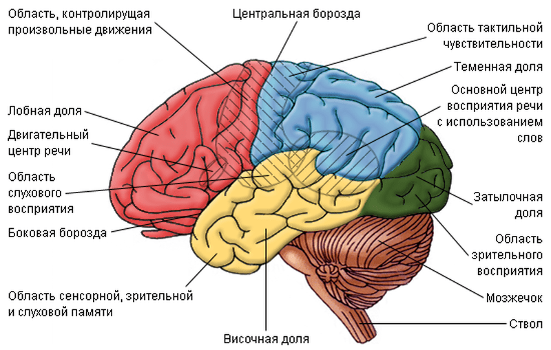
BRAIN consists of:
two hemispheres
cerebrum brainstem,
cerebellum.
Hemispheres of the brain divided into frontal lobes, parietal, temporal and occipital lobes. The hemispheres of the brain are connected through the corpus callosum.
- The frontal lobes are responsible for the intellectual and emotional sphere, thinking and complex behavior, conscious movements, motor speech and writing skills.
- Temporal lobes are responsible for hearing, sound perception, vestibular information, partial analysis of visual information (for example, face recognition), sensory part of speech, participation in memory formation, influence on the emotional background, for influence on the autonomic nervous system through communication with the limbic system.
- The parietal lobes are responsible for various types of sensitivity (tactile, pain temperature, deep and complex spatial types of sensitivity), spatial orientation and spatial skills, reading, counting.
- Occipital lobes - perception and analysis of visual information.
brain stem represented by the diencephalon (thalamus, epithalamus, hypothalamus and pituitary), midbrain, pons and medulla oblongata. Functions of the brain stem responsible for unconditioned reflexes, influence on the extrapyramidal system, taste, visual, auditory and vestibular reflexes, suprasegmental level of the autonomic system, control of the endocrine system, regulation of homeostasis, hunger and satiety, thirst, regulation of the sleep-wake cycle, regulation of respiration and the cardiovascular system , thermoregulation.
Cerebellum consists of two hemispheres and a worm that connects the hemispheres of the cerebellum. Both the cerebral hemispheres and the cerebellar hemispheres are striated with furrows and convolutions. The cerebellar hemispheres also contain nuclei with gray matter. The cerebellar hemispheres are responsible for coordination of movements and vestibular function, and the cerebellar vermis is responsible for maintaining balance and postures, muscle tone. The cerebellum also influences the autonomic nervous system. There are four ventricles in the brain, in the system of which CSF circulates and which are connected with the subarachnoid space of the cranial cavity and spinal canal.

Spinal cord consists of the cervical, thoracic, lumbar and sacral regions, has two thickenings: the cervical and lumbar, and the central spinal canal (in which the cerebrospinal fluid circulates and which in the upper sections connects to the fourth ventricle of the brain).
Histologically, brain tissues can be divided into Gray matter, which contains neurons, dendrites (short processes of neurons) and glial cells, and white matter, in which axons lie, long processes of neurons covered with myelin. In the brain, gray matter is located mainly in the cerebral cortex, in the basal nuclei of the hemispheres and the nuclei of the brain stem (midbrain, bridge and medulla oblongata), and in the spinal cord, gray matter is located in depth (in its central sections), and the outer parts of the spinal cord are represented by white matter.
Peripheral nerves can be divided into motor and sensory, forming reflex arcs that are controlled by parts of the central nervous system.

autonomic nervous system has a division into suprasegmental and segmental.
- The suprasegmental nervous system is located in the limbic-reticular complex (structures of the brain stem, hypothalamus and limbic system).
- The segmental part of the nervous system is divided into the sympathetic, parasympathetic and metasympathetic nervous systems. The sympathetic and parasympathetic nervous systems are also divided into central and peripheral. The central divisions of the parasympathetic nervous system are located in the midbrain and medulla oblongata, and the central divisions of the sympathetic nervous system are located in the spinal cord. The metasympathetic nervous system is organized by nerve plexuses and ganglia in the walls of the internal organs of the chest (heart) and abdominal cavity (intestines, bladder, etc.).
The human nervous system is the main link that binds together all human organs and forms their inseparable activity. The nervous system, namely the brain, is the organ that creates our psyche. All our thoughts, experiences, behavior, emotions, memory arise under the influence of electrochemical reactions occurring between neurons - nerve cells.
The brain has a huge number of neurons, and even more - their connections. Therefore, it is not in vain that it is compared with the Universe, since the possibilities of this organ are almost limitless.
The structure of the human nervous system
The nervous system consists of two anatomical sections - central (CNS) and peripheral (PNS).
The CNS includes the brain and spinal cord. The main role in the transmission and generation of electrical impulses is played by the brain, and the spinal cord is the link between it and the nerves that form the peripheral nervous system.
Functions of the human nervous system
Both departments work in harmony. For example, if we touch a rough surface, receptors on the skin transmit a signal through the peripheral nerves, which form nerve bundles, and are sent to the thoracic spinal cord. The latter redirects the signal to the brain. It reaches the area of the cortex responsible for tactile sensitivity. Thanks to this, we evaluate the surface that we touch.
If we want to do something with our own hands, for example, move a pen from one place to another, the following happens. The region of the cerebral cortex responsible for the contraction of the muscles of the upper limbs gives a signal to the underlying structures, which is transmitted to the spinal cord. The latter sends an impulse to the thoracic plexus. Further, the signal goes to the hands along the ulnar and radial nerves, moving the muscles.

It should be said that not all actions take place with the participation of the brain. For example, unconditioned reflexes close at the level of the spinal cord. So, if we touch a hot surface, the impulse goes to the sensitive neurons of the spinal cord, which communicate with the motor cells, and they send a signal to the muscles of the hands so that we react to the danger as quickly as possible and not be injured.
We also owe the relative autonomy of the spinal cord to our ability to ride a bicycle, dance, perform subtle physical movements, and even walk. When a person first encounters these activities, the brain is actively involved in the formation of reflexes. Therefore, we think about every movement, every subtlety (pedal pressure, foot placement, etc.). Then the reflex is fully formed, reaching automatism, after which its implementation no longer requires our attention. After all, the cyclist does not think about which foot to pedal, and an adult (unlike a baby) does not follow every position of his foot when walking.
The human autonomic nervous system
But most of the physiological functions take place without our participation at all. For this there is an autonomic nervous system. This is a combination of nerves and their plexuses, which ensures the functioning of internal organs (heart, lungs, intestines). Therefore, we do not need to think that we need to breathe or digest food, we do not need to forcefully form a peristalsis wave so that nutrients pass through the intestines. All this is done by the autonomic nervous system, which is represented by both nerves extending from the spinal cord and cranial nerves that begin in the brain stem and subcortical structures.
The brain is the "administrator" of the nervous system and the human psyche
Previously, it was believed that the “soul” of a person is enclosed in his heart. With the development of science, humanity gradually began to study what made us people, the crown of the animal world - the brain.
Our mental system is formed due to the interaction of the cerebral cortex - and the underlying sections (intermediate, midbrain, trunk). Each area is responsible for a particular function. But the most interesting thing is that when one section of neurons fails, its work can be partially replaced by another, which is called neuroplasticity.
The frontal lobe is involved in the formation of emotions, memory, speech and behavior. Evolutionarily, this part of Homo sapiens is the most developed, since it began to develop with the transition of primates to upright posture and the activation of fine motor skills of the upper limbs. Therefore, the frontal lobe is responsible for many functions. To understand what effect the frontal lobe has on the mental state of a person, we should mention the so-called frontal syndrome, which is observed in people with organic brain damage due to external influences, vascular, oncological and other pathologies. They have a disinhibition in behavior, self-control disappears and a tendency to brutal, antisocial acts, as well as outbursts of aggressiveness, appears. In addition to behavior and emotions, memory is disturbed, a person cannot concentrate on some business, the function of cognition of the external world suffers. In severe cases, the core of the personality is lost - we stop seeing the person as he was before.
There is a part of the brain called hypothalamus, which is responsible for the regulation of vegetative functions. It communicates with the endocrine system of the body and is directly related to the gland-regulator of hormones - pituitary gland. The latter secretes special substances that give a signal to the pituitary gland to release hormones: gonadotropic (affecting the sex glands), thyrotropic (on the thyroid gland), adrenocorticotropic (on the adrenal glands), somatotropic (on tissue growth) and prolactin (on the mammary glands).
Biochemistry of the nervous system and human psyche
To connect neurons with each other, there are biologically active substances responsible for the connection between neurons - neurotransmitters, each of which has its own function.

Consider the main types of neurotransmitters:
Neurotransmitters act by passing through synapses in neurons - connections between cells. Substances pass through the synaptic cleft - the space between neurons - stimulating receptors and forming an electrical impulse, which is the means of signal transmission in the nervous system.
Thus, the nervous system is responsible not only for the formation of the psyche, but also controls the somatic sphere of the human body. And the brain is the main “administrator” here.
The human brain is one of the most unsolved mysteries. And we still have a lot to learn about ourselves by studying it.
 Two heads and six legs; four walk, and two lie still
 Self-esteem - what is it: concept, structure, types and levels
 Cassandra's Path, or Pasta Adventures War on Earth and Underground