Open lesson in biology using ICT "History and methods of studying the cell. Cell theory" (9th grade)
A kind of "building block" of living nature is a cell - an elementary, but rather complex biosystem. Eukaryotic cells are extremely diverse.
1) What does cytology study? What do you know about the development of the science of cytology? What is the role of instruments in the study of the cell?
- Answer: Cytology studies the composition, structure and function of cells. Devices, in particular a microscope, play an important role in the development of cytology, as they allow us to examine the structure of cells.
2) Among the cells listed below, write out those that: a) are part of a multicellular organism; b) are free-living organisms.
3) Complete the definition.
- Answer: Cells that are similar in structure, located side by side, united by an intercellular substance and performing certain functions, form tissues and organs. Tissues of plants and animals that perform protective functions are called protective (skin) cover.
4) You know that animals have four types of tissues. What type are:
b) Top layer of skin
c) brain cells
d) rectus dorsi muscle
e) Olfactory cells of the nasal cavity
e) subcutaneous tissue
g) Platelet
a) connective
b) Epithelial
c) nervous
d) Muscular
e) nervous
f) Connecting
g) Blood cells (connective)
5) What types of fabrics are typical for:
a) an apple leaf
b) maple bark
c) pine needles
d) potato tubers
e) poplar trunk?
a) Apple leaf: the epidermis is covered on the outside - the integumentary tissue, between the upper and lower epidermis is an assimilation parenchyma or chlorenchyma, which belongs to the main tissues. Leaf veins are vascular bundles, which include vascular elements - xylem and phloem. The bundles are surrounded by mechanical tissue.
b) Maple bark consists of conducting elements of the phloem, the main parenchyma, and bast fibers. If you mean the outer cover - this is the crust, which is a collection of several periderms - the integumentary tissue.
c) Pine needles have the same structure as the leaves of an apple tree. The only difference is that under the epidermis there is a hypodermis (also integumentary tissue), folded chlorenchyma and there are resin passages
e) Poplar trunk: on the outside, the integumentary tissue is the periderm or crust, then comes the bark, which has the same structure as the maple bark, then the cambium (educational tissue). The composition of wood includes conductive tissue (xylem), the main parenchyma (axial and radial) and wood fibers (mechanical tissue). In the center, the core consists of cells of the main parenchyma.
6) Prove that a cell is a structural and functional unit of living organisms.
- Answer: A cell is capable of reproducing a similar cell to itself.
7) Describe how knowledge about the cell developed. Mark the main stages in the development of cytological knowledge.
- Answer: From the creation of the microscope, then the discovery of the cell, later the creation of the cell theory, and then the discovery of the smallest organelles, biochemistry and, of course, genetic discoveries.
8) Carefully read the main provisions of the cell theory in the textbook, mentally mark them with numbers. Please indicate to which of these statements the following statements may apply:
a) plant cells contain carbohydrates and various mineral salts
b) root growth depends on the functioning of a certain zone of it
c) plants, animals, fungi and all living things on our planet are made up of cells
- Answer: All living things are made up of cells (exception: viruses). All physiological processes take place at the level of the cell, then at the level of the organ and organism. Any pathological process is based on changes in the functions and structure of cells. Division occurs at the level of cells, growth is an increase in the number of cells.

Hooke Robert 1635 - 1703 Robert Hooke - English naturalist was born on the Isle of Wight in the family of a local church priest. His father initially prepared him for spiritual activity, but then, in view of the boy’s poor health and his ability to engage in mechanics, he assigned him to study watchmaking. Subsequently, however, young Robert showed an interest in scientific studies, and as a result was sent to Westminster School, where he successfully studied Latin, Greek, Hebrew, but was especially interested in mathematics and showed a great ability for inventions in physics and mechanics. His ability to study physics and chemistry was recognized and appreciated by scientists at Oxford University, where he began to study in 1653. First he was an assistant to the chemist Willis, and then to the famous Boyle. During his 87 years of life, Robert Hooke, despite poor health, was tireless in his studies, made many scientific discoveries, inventions and improvements. In 1663 he was appointed curator of experiments at the newly founded Royal Society of London. Since 1665 - professor at the University of London, in 1677-1683. - Secretary of the Royal Society of London. With the help of a microscope he improved, Hooke observed the structure of plants and gave a clear drawing, which for the first time showed the cellular structure of cork. The term "cell" was first introduced by Hooke. In his work "Micrography", published in 1665, he described the cells of elder, dill, carrots, gave images of very small objects, such as the eye of a fly, a mosquito and its larvae, described in detail the cellular structure of cork, bee wing, mold, moss. In the same work, Hooke outlined his theory of colors, he adhered to the wave theory of light and challenged the corpuscular, considered heat to be the result of the mechanical movement of particles of matter. Hooke expressed thoughts about the change in the earth's surface, which, in his opinion, led to a change in the fauna. He believed that fossils are the remains of previously living creatures, from which the history of the Earth can be reproduced. Hook was also known as an architect. Several buildings were built according to his designs, mainly in London.

Anthony van Leeuwenhoek Anthony van Leeuwenhoek (Leeuwenhoek) () is a Dutch naturalist, one of the founders of scientific microscopy. Having made lenses with multiple magnification, for the first time he observed and sketched (publications since 1673) a number of protozoa, spermatozoa, bacteria, erythrocytes and their movement in capillaries. Anthony van Leeuwenhoek has been improving his microscopes all his life: he changed lenses, invented some devices, varied the conditions of the experiment. For many years Leeuwenhoek made his lenses in the form of lentils, called "microscopy", the lenses were essentially magnifiers. They were tiny, sometimes smaller than a fingernail, but magnified by 100 and even 300 times. To observe with these lenses, it was necessary to acquire certain skills and be patient. There is no data to establish with accuracy when Leeuwenhoek began research. He was far from thinking of making a discovery: for him, an adult and respectable person, the microscope was just a favorite toy. But it was impossible to break away. After his death, 273 microscopes and 172 lenses were counted in his office, which he called a museum, 160 microscopes were mounted in silver frames, 3 in gold. And how many devices he lost because he tried, at the risk of his own eyes, to observe under a microscope the moment of the explosion of gunpowder.

Schwann Theodor 1810–1882 Theodor Schwann was the first scientist to establish that the cell is the microscopic element that makes up all living tissues, all organs and all microscopic living beings. Schwann came to the conclusion that plants and animals develop on the same basis and that the law of cell structure is the same for them. In 1839, Schwann published his work "Microscopic studies on the conformity in the structure and growth of animals and plants." Labor caused a revolution in biology. Thus, one of the most important biological theories, called the cell theory, was developed. Theodor Schwann was born in Neuss on December 7, 1810. After graduating (in 1833) from the University of Bonn and after studying in Cologne and Würzburg, he entered the Berlin Anatomical Institute. In 1834–1838, while working as an assistant, Schwan made a number of scientific discoveries. He established the cellular structure of the dorsal chord, the walls of blood vessels, muscles, cartilage, etc. In 1838 he gave a description of a peculiar thin sheath surrounding the peripheral nerve fibers, called the Schwann sheath, in the same year he published three reports on these topics, which included in his main work, published in 1839. In this work, the scientist proved the cellular theory of the structure of organisms. He put several premises at the basis of this theory: both plants and animals are characterized by unity of structure; the basis of the structure of all organisms is a cell; the formation of more and more new cells is the principle of organic growth and development of plants and animals; a cell is an elementary biological unit; The organism as a whole is the sum of the cells that formed it. On the basis of cell theory, it finally became clear that the fruit membranes grow and form folds by gradually increasing the number of cells that are arranged in a certain way. The egg and sperm are just separate germ cells. As soon as they unite, all new individual cells begin to appear, from which the germ (embryo) of the corresponding organism then arises. Theodor Schwann died in Cologne on January 14, 1882.

Schleiden Matthias Jakob 1804–1881 Together with the zoologist Theodor Schwann, Schleiden engaged in microscopic research, which led scientists to develop a cellular theory of the structure of organisms. In 1839 Schleiden received his Ph.D. from the University of Jena. He received his doctorate in medicine in 1843 at the University of Tübingen, and from 1863 he was a professor of photochemistry (the science of chemical processes in living plants) and anthropology in Dorpat, and also conducted scientific work in Dresden, Wiesbaden and Frankfurt. In the book Data on Phylogeny, in the section on the origin of plants, Schleiden presented his theory of the origin of the offspring of cells from the mother cell. Schleiden's work prompted Theodor Schwann to engage in lengthy and thorough microscopic studies that proved the unity of the cellular structure of the entire organic world. The work of the scientist under the title "The Plant and Its Life" was published in 1850 in Leipzig. Schleiden's main work, Fundamentals of Scientific Botany, was published in two volumes in Leipzig and had a tremendous impact on the reform of plant morphology based on ontogeny. Ontogeny distinguishes three periods in the development of an individual organism: the formation of germ cells; the body, the embryonic period, which is limited to the formation of eggs and spermatozoa; the embryonic period - from the beginning of the division of the egg to the birth of the individual; postpartum period - from the birth of an individual to his death. At the end of his life, Schleiden left botany and took up anthropology, i.e. the science of differences in appearance, structure and activity of organisms of individual human groups in time and space. Schleiden died on June 23, 1881 in Frankfurt am Main.

Ilya Ilyich Mechnikov Ilya Ilyich Mechnikov () Russian biologist and pathologist, one of the founders of comparative pathology, evolutionary embryology and domestic microbiology, immunology, creator of the theory of phagocytosis and the theory of immunity, founder of a scientific school, corresponding member (1883), honorary member (1902) ) Petersburg Academy of Sciences. He discovered in 1882 the phenomenon of phagocytosis. In the works "Immunity in infectious diseases" (in 1901) he outlined the phagocytic theory of immunity. Created the theory of the origin of multicellular organisms. Proceedings on the problem of aging. Nobel Prize (1908, jointly with the German physician, bacteriologist and biochemist Paul Ehrlich).

Navashin Sergey Gavrilovich In 1898 he discovered double fertilization in angiosperms. He laid the foundations of chromosome morphology and karyosystematics. Author of a number of works on mycology and comparative anatomy

Modern cell theory includes the following provisions: * cell - the basic unit of the structure and development of all living organisms, the smallest unit of life; * cells of all unicellular and multicellular organisms are similar (homologous) in their structure, chemical composition, basic manifestations of vital activity and metabolism; * cell reproduction occurs by dividing, and each new cell is formed as a result of the division of the original (mother) cell; * in complex multicellular organisms, cells are specialized in their function and form tissues; tissues consist of organs that are closely interconnected and subordinate to the nervous and humoral systems of regulation.

General characteristics of cells Cells of plant and animal tissues have different shapes and sizes depending on the functions they perform. The diameter of most cells ranges from 10 to 100 microns. The smallest cells are about 4 µm in size. However, there are also very large cells visible to the naked eye (cells of the pulp of watermelon, eggs). The shape of the cell can be round, polygonal, rod-shaped, stellate, process, cylindrical, cubic, etc. The cell is an elementary living system, consisting of three main structural elements - membranes, cytoplasm and nuclei. The cytoplasm and nucleus form protoplasm.


Properties of the cell The cell consists of a jelly-like mass - protoplasm and nucleus, surrounded by a cell membrane. Cells have all the properties of living matter, including self-preservation and self-reproduction. absorption and assimilation. Cells selectively absorb chemicals such as amino acids from the surrounding intercellular (interstitial) fluid, from which more complex compounds are synthesized - proteins that form the basis of protoplasm. Thus, the cell is a unit that actively accumulates and uses nutrients that enter the human body with food. Growth and recovery. Nutrients can be used to synthesize new protoplasm, resulting in an increase in size. In addition, nutrients are necessary for the restoration (regeneration) of parts of cells that have become unusable. Metabolism. Growth and regeneration are carried out due to the anabolic function, for which the cell needs energy. As its source, individual components of substances entering the cell are used. The energy released during their splitting (catabolism) is necessary for the cell for heat production, release of secrets, movements and nervous activity. Breath. For the functioning and maintenance of the activity of the cell, the delivery of oxygen from the lungs with the blood flow and the removal of carbon dioxide (the end product of metabolism) from the tissues are essential. Selection. The substances formed as a result of catabolic processes are released from the cell into the interstitial fluid, from where they enter the blood. In this case, carbon dioxide is transported to the lungs and removed from the body in the form of carbon dioxide. Other metabolic products are excreted through the kidneys in the urine.

Internet resources c4c7d84/85238/lesson c4c7d84/85238/lesson %D0%BA,_%D0%90%D0%BD%D1%82%D0%BE%D0%BD%D0%B8_%D0%B2%D0%B0% D0%BD %D0%BA,_%D0%90%D0%BD%D1%82%D0%BE%D0%BD%D0%B8_%D0%B2%D0%B0%D0%BD html html htm htm html

Sections: Biology
Class: 9
Target: to introduce students to the history of the discovery and study of the cell, the main provisions of the cell theory and methods of studying the cell.
Educational tasks: to form knowledge about the history of the discovery of the cell, scientists who have contributed to the study of the structure of the cell; to analyze the main provisions of the cell theory; to find out what methods are used in cytology to study the cell.
Development tasks: continue the formation of skills and abilities to use various information sources: the Internet, additional literature in preparation for the lesson, the ability to compare, analyze, draw conclusions, develop students' monologue speech, improving the technique of public speaking.
Educational tasks: the formation of independent work skills to show the general biological significance of cell theory.
Method: reproductive.
Equipment: multimedia projector, smart board, table "General scheme of the structure of plant and animal cells."
During the classes
1. Organizational and psychological moment
Welcoming children by the teacher, friendly atmosphere.
Announcement of the topic of the lesson by the teacher (it is written on the board in advance).
In the notebook, the children write the topic of the lesson.
Joint definition with students of the goals and objectives of the lesson.
Set up productive collaboration.
2. Knowledge check
Task 1. Blitz survey. The teacher asks questions, the answers should be precise and short.
- What are the kingdoms of wildlife? (plants, animals, mushrooms, plants)
- What are the living kingdoms made of? (cells)
- What other fifth kingdom exists? (viruses)
- What is the nature of viruses? (have no cellular structure)
- What are the levels of organization of life? (molecular, cellular, organismal, population-species, biogeocenotic, biospheric)
Task 2. Charging for the mind.
Teacher. We have studied the signs of living organisms. Only a few are written on the board, only the letters are all mixed up. Find and write down these signs in your notebook. The first three who answer correctly will receive excellent marks:
- liquidmoraz - irritability;
- taniyepi - food;
- guciasaremolya - self-regulation;
- variety - growth;
- vitierase - development;
- ice-coveredness - heredity;
- chaneids - breath;
- multiple - reproduction.
Task 3. Remember, compare, show. Teacher. On the board hangs a table "The structure of plant and animal cells." What unites these cells? Go to the board and on the table show the common components (nucleus, membrane, cytoplasm)
3. New material
(introductory speech of the teacher)
The cell is an interesting, amazing and mysterious world that is characteristic of any living creature, excluding viruses. But it was possible to solve the secrets of the cell only with the invention of the first microscope at the end of the 16th century. The history of the study of the cell is inextricably linked with the development of microscopic techniques and research methods. The invention of the microscope led to an in-depth study of the organic world.
In 1590, the Dutchman Hans Jansen designed the first optical instrument, which consisted of a tube with two magnifying glasses attached to a stand.
Slides 2-4. In 1665, the English naturalist Robert Hooke, examining a section of cork oak bark under a microscope improved by him, saw formations resembling a honeycomb. Describing what he saw, Hooke used the word "kel", which in English means "chamber", "cell". The term was translated into Russian as CELL.
Therefore, we use the term cell thanks to Robert Hooke.
Although now we know that he saw not the cells themselves, but their cell walls.
Slide 5. In the period from 1676 to 1719, Hooke's contemporary, the Dutch merchant Anthony van Leeuwenhoek, won the fame of a scientist and presented science with the greatest discovery. He improved Hooke's microscope and created lenses that give a magnification of 100-300 times and discovered the world of unicellular organisms.
Leeuwenhoek wrote “Oh eureka! People that I see! In this small drop of water I met a whole world of small living beings. A world that is difficult to understand and explain. These little animals were very funny, they tumbled, jumped, frolicked and were very happy in life.
Yes, and in the form of "animals" were quite cute: balls, spirals, sticks, then one by one, then 2-3 in a dance that was understandable only to them.
The descriptions of these "little animals" earned the Dutchman worldwide fame, prompted an interest in the study of the living world.
Slide 6. In 1831, the Scottish botanist Robert Brown first described the nucleus in a plant cell.
In 1834, the Russian scientist P.F. Goryaninov in his research noted that all animals and plants consist of interconnected cells, which he called vesicles, sacs or closets. He expressed his opinion on the general plan of the structure of plants and animals.
In the middle of the 19th century, a lot of information and new knowledge about cells accumulated.
Slide 7. 1838 German botanist Matthias Schleiden came to the conclusion that plant tissues are made up of cells.
Slide 8. 1839 The German physiologist Theodor Schwann published the book "Microscopic studies on the correspondence in the structure and growth of animals and plants", in which he formulated the conclusion that the cell is the structural and functional unit of living organisms.
This idea is called the Schwann-Schleiden theory.
Slide 9. The main provisions of the Schwann-Schleiden theory (1838-1839):
- All organisms are made up of cells.
- Cells are the smallest structural units of life.
- Cells in the body arise by neoplasms from non-cellular substances.
Slide 10. Scientists' mistakes.
M. Schleiden and T. Schwann erroneously believed that cells arise by neoplasm from non-cellular substance.
Slide 11. Karl Baer, an academician of the Russian Academy in 1827, discovered the egg cell of mammals. Baer established that all organisms begin their development from a single cell (zygote). This discovery proves that the cell is also a unit of development of all living organisms.
Slide 12. 1840 Jan Purkyne proposed the term "protoplasm" to refer to the living contents of the cell. 1844. The scientist Hugo Mol described the contents of the cell in detail using the term "protoplasm".
Slide 13. 1855 The German physician Rudolf Virchow convincingly proved that cells arise only from cells, by reproduction - “cells from a cell”, refuting the erroneous idea of Schleiden and Schwann's cell formation. Virchow's mistake was that he believed that cells are weakly connected with each other and exist each on its own. Later it was possible to prove the integrity of the cellular system.
Slide 14. 1876 Alexander Fleming discovered the cell center.
Slide 15. 1890 Richard Altmann discovered mitochondria.
Slide 15. 1898 Camillo Golgi discovered an organoid named after him - the Golgi apparatus or complex.
Slide 16. At the turn of the 19th and 20th centuries, a new biological science, CYTOLOGY, was formed (from the Greek cytos - cell, logos - teaching)
CYTOLOGY studies:
- The structure of cells and their organelles;
- Functions of organelles and other cell structures;
- Reproduction and development of cells.
slide 17. MAIN PROVISIONS OF MODERN CELL THEORY
- The cell is a structural and functional unit of the living, which is an elementary living system. It is characterized by all the signs of a living thing.
- Slide 18. The cells of all organisms have a similar chemical composition and general structural plan.
- Slide 19. A new cell arises as a result of the division of the original cell.
- Slide 20. Multicellular organisms develop from a single source cell.
- The similarity of the cellular structure indicates the unity of their origin.
Slide 21. CELL STUDY METHODS
MICROSCOPY:
- LIGHT MICROSCOPE. Studies cellular forms and structures: nucleus, mitochondria, chloroplasts, Golgi apparatus.
- Slide 22. ELECTRONIC MICROSCOPE. Invented in the 30s of the 20th century. Modern microscopes provide magnification up to 1,000,000 times. It allows you to consider the structure of cell organelles in more detail.
- Slide 23. DIFFERENTIAL CENTRIFUGATION METHOD. It is based on the different densities of organoids and, with very fast rotation on a centrifuge, organelles are arranged in layers in solution in accordance with their density.
- Slide 24. FLUORESCENT MICROSCOPY. Living cells are observed in ultraviolet light. At the same time, some components begin to glow immediately, while others glow when special dyes are added. Fluorescent microscopy allows you to see the location of nucleic acids, vitamins, fats.
- CELL AND TISSUE CULTURE METHOD. It allows you to see the growth of cells, to observe reproduction, to determine the effect of various substances on cells, to obtain cell hybrids.
Slide 25. THE SIGNIFICANCE OF CELL STUDIES:
- In medicine, to unravel the causes of diseases.
- To classify living organisms.
- In genetics (hereditary diseases, mutations).
- In agriculture (genetic, cell engineering, selection).
- To unlock the mysteries of evolution.
Slide 26. CONCLUSION:
- The cell is the structural unit of all living organisms (except viruses).
- The commonality of the chemical composition and structure speaks of the unity of the origin of all life on Earth.
4. FIXING
And now we will check how you understood the material of today's lesson?
Slide 27. TASK 1
Complete the test work in your notebook and rate yourself.
1. For the first time I saw and described plant cells:
- R. Virchow;
- R. Hooke;
- K. Baer;
- A. Leeuwenhoek.
2. Improved the microscope and saw single-celled organisms for the first time:
- M. Schleiden;
- A. Levenguk;
- R. Virchow;
- R. Hooke.
3. The creators of the cell theory are:
- Ch. Darwin and A. Wallace;
- T. Schwann and M. Schleiden;
- G. Mendel and T. Morgan;
- R. Hooke and N. G.
4. The cell theory is unacceptable for:
- fungi and bacteria;
- viruses and bacteria;
- animals and plants;
- bacteria and plants.
5. The cellular structure of all living organisms indicates:
- unity of chemical composition;
- variety of living organisms;
- the unity of the origin of all living things;
- unity of animate and inanimate nature.
Now let's look at the correct answers.
slide 28. ANSWERS:
Correct the mistakes and rate according to the criteria:
5 - everything is correct;
4 - one mistake;
3 - two errors;
2 - three mistakes.
Teacher: please raise your hands who received "5", "4", "3", "2" (everything counts and announces the result).
5. Homework
Paragraph 10, using Cicero's verbal formula: Who? What? Where? How? For what? How? When? Make up questions for the paragraph.
slide 2
From the history of cytology
slide 3
Hooke Robert 1635 - 1703
- Robert Hooke - English naturalist was born on the Isle of Wight in the family of a local church priest. His father initially prepared him for spiritual activity, but then, in view of the boy’s poor health and his ability to engage in mechanics, he assigned him to study watchmaking. Subsequently, however, young Robert showed an interest in scientific studies, and as a result was sent to Westminster School, where he successfully studied Latin, Greek, Hebrew, but was especially interested in mathematics and showed a great ability for inventions in physics and mechanics. His ability to study physics and chemistry was recognized and appreciated by scientists at Oxford University, where he began to study in 1653. First he was an assistant to the chemist Willis, and then to the famous Boyle. During his 87 years of life, Robert Hooke, despite poor health, was tireless in his studies, made many scientific discoveries, inventions and improvements. In 1663 he was appointed curator of experiments at the newly founded Royal Society of London. Since 1665 - professor at the University of London, in 1677-1683. - Secretary of the Royal Society of London.
- With the help of a microscope he improved, Hooke observed the structure of plants and gave a clear drawing, which for the first time showed the cellular structure of cork. The term "cell" was first introduced by Hooke. In his work "Micrography", published in 1665, he described the cells of elder, dill, carrots, gave images of very small objects, such as the eye of a fly, a mosquito and its larvae, described in detail the cellular structure of cork, bee wing, mold, moss. In the same work, Hooke outlined his theory of colors, he adhered to the wave theory of light and challenged the corpuscular, considered heat to be the result of the mechanical movement of particles of matter. Hooke expressed thoughts about the change in the earth's surface, which, in his opinion, led to a change in the fauna. He believed that fossils are the remains of previously living creatures, from which the history of the Earth can be reproduced. Hook was also known as an architect. Several buildings were built according to his designs, mainly in London.
slide 4
Anthony van Leeuwenhoek 1632-1723
- Anthony van Leeuwenhoek (1632-1723) - Dutch naturalist, one of the founders of scientific microscopy. Having made lenses with 150-300-fold magnification, for the first time he observed and sketched (publications since 1673) a number of protozoa, spermatozoa, bacteria, erythrocytes and their movement in capillaries. Anthony van Leeuwenhoek has been improving his microscopes all his life: he changed lenses, invented some devices, varied the conditions of the experiment.
- For many years Leeuwenhoek made his lenses in the form of lentils, called "microscopy", the lenses were essentially magnifiers. They were tiny, sometimes smaller than a fingernail, but magnified by 100 and even 300 times. To observe with these lenses, it was necessary to acquire certain skills and be patient. There is no data to establish with accuracy when Leeuwenhoek began research. He was far from thinking of making a discovery: for him, an adult and respectable person, the microscope was just a favorite toy. But it was impossible to break away. After his death, 273 microscopes and 172 lenses were counted in his office, which he called a museum, 160 microscopes were mounted in silver frames, 3 in gold. And how many devices he lost - after all, he tried, at the risk of his own eyes, to observe under a microscope the moment of the explosion of gunpowder.
slide 5
Schwann Theodor 1810–1882
- Theodor Schwann was the first scientist to establish that the cell is the microscopic element that makes up all living tissues, all organs and all microscopic living beings.
- Schwann came to the conclusion that plants and animals develop on the same basis and that the law of cell structure is the same for them. In 1839, Schwann published his work "Microscopic studies on the conformity in the structure and growth of animals and plants."
- Labor caused a revolution in biology. Thus, one of the most important biological theories, called the cell theory, was developed.
- Theodor Schwann was born in Neuss on December 7, 1810. After graduating (in 1833) from the University of Bonn and after studying in Cologne and Würzburg, he entered the Berlin Anatomical Institute. In 1834–1838, while working as an assistant, Schwan made a number of scientific discoveries.
- He established the cellular structure of the dorsal chord, the walls of blood vessels, muscles, cartilage, etc. In 1838 he gave a description of a peculiar thin sheath surrounding the peripheral nerve fibers, called the Schwann sheath, in the same year he published three reports on these topics, which included in his main work, published in 1839. In this work, the scientist proved the cellular theory of the structure of organisms. He based this theory on several premises:
- both plants and animals are characterized by unity of structure;
- the basis of the structure of all organisms is a cell;
- the formation of more and more new cells is the principle of organic growth and development of plants and animals;
- a cell is an elementary biological unit;
- The organism as a whole is the sum of the cells that formed it.
- On the basis of cell theory, it finally became clear that the fruit membranes grow and form folds by gradually increasing the number of cells that are arranged in a certain way. The egg and sperm are just separate germ cells. As soon as they unite, all new individual cells begin to appear, from which the germ (embryo) of the corresponding organism then arises. Theodor Schwann died in Cologne on January 14, 1882.
slide 6
Schleiden Matthias Jakob 1804–1881
- Together with the zoologist Theodor Schwann, Schleiden engaged in microscopic research, which led scientists to develop a cellular theory of the structure of organisms. In 1839 Schleiden received his Ph.D. from the University of Jena. He received his doctorate in medicine in 1843 at the University of Tübingen, and from 1863 he was a professor of phytochemistry (the science of chemical processes in living plants) and anthropology in Dorpat, and also conducted scientific work in Dresden, Wiesbaden and Frankfurt.
- In the book "Data on Phytogenesis" in the section on the origin of plants, Schleiden presented his theory of the origin of the offspring of cells from the mother cell. Schleiden's work prompted Theodor Schwann to engage in lengthy and thorough microscopic studies that proved the unity of the cellular structure of the entire organic world. The work of the scientist under the title "The Plant and Its Life" was published in 1850 in Leipzig.
- Schleiden's main work "Fundamentals of Scientific Botany" in two volumes was published in 1842-1843 in Leipzig and had a huge impact on the reform of plant morphology based on ontogeny. Ontogeny distinguishes three periods in the development of an individual organism:
- the formation of germ cells, i.e. the pre-embryonic period, limited by the formation of eggs and spermatozoa;
- the embryonic period - from the beginning of the division of the egg to the birth of the individual;
- postpartum period - from the birth of an individual to his death.
- At the end of his life, Schleiden left botany and took up anthropology, i.e. the science of differences in appearance, structure and activity of organisms of individual human groups in time and space.
- Schleiden died on June 23, 1881 in Frankfurt am Main.
Slide 7
Ilya Ilyich Mechnikov 1845-1916
Ilya Ilyich Mechnikov (1845-1916) - Russian biologist and pathologist, one of the founders of comparative pathology, evolutionary embryology and domestic microbiology, immunology, creator of the theory of phagocytosis and the theory of immunity, creator of a scientific school, corresponding member (1883), honorary member ( 1902) Petersburg Academy of Sciences. He discovered in 1882 the phenomenon of phagocytosis. In the works "Immunity in infectious diseases" (in 1901) he outlined the phagocytic theory of immunity. Created the theory of the origin of multicellular organisms. Proceedings on the problem of aging. Nobel Prize (1908, jointly with the German physician, bacteriologist and biochemist Paul Ehrlich).
Slide 8
Navashin Sergey Gavrilovich 12/14/1857 - 12/10/1930
- In 1898 he discovered double fertilization in angiosperms.
- He laid the foundations of chromosome morphology and karyosystematics. Author of a number of works on mycology and comparative anatomy.
Slide 9
Modern cell theory includes the following provisions
- cell - the basic unit of the structure and development of all living organisms, the smallest unit of life;
- cells of all unicellular and multicellular organisms are similar (homologous) in their structure, chemical composition, basic manifestations of vital activity and metabolism;
- cell reproduction occurs by dividing, and each new cell is formed as a result of the division of the original (mother) cell;
- in complex multicellular organisms, cells are specialized in their function and form tissues; tissues consist of organs that are closely interconnected and subordinate to the nervous and humoral systems of regulation.
Slide 10
General characteristics of cells
Plant and animal tissue cells have different shapes and sizes depending on the functions they perform. The diameter of most cells ranges from 10 to 100 microns. The smallest cells are about 4 µm in size. However, there are also very large cells visible to the naked eye (cells of the pulp of watermelon, eggs). The shape of the cell can be round, polygonal, rod-shaped, stellate, process, cylindrical, cubic, etc. The cell is an elementary living system, consisting of three main structural elements - membranes, cytoplasm and nuclei. The cytoplasm and nucleus form protoplasm.
slide 11
bacteria
slide 12
Diversity of euraryotic cells
LESSON 4. CYTOLOGY - THE SCIENCE OF THE CELL. VARIETY OF CELLS. PURPOSE OF THE LESSON: to recall the history of the development of cytology; consider the diversity of cells that exist in wildlife LESSON OBJECTIVES: Get acquainted with the history of the development of the science of cells - cytology Consider the diversity of cells in connection with their functions EQUIPMENT: tables "Plant cell and animals", "Eukaryotic and prokaryotic cells", cells"; electronic presentations - Presentation No. 1 "History of the development of cytology", Presentation No. 2 "Diversity of cells"; microscopes and micropreparations of cells. LESSON PROCESS: Slide 1 - Today we are moving on to studying one of the most important, but at the same time very complex sections of general biology - to studying the basics of cytology. After all, one of the fundamental concepts in biology is the idea that the structural and functional unit of living organisms is a cell. Slide 2 - Dictionary Cytology (Greek κύτος - bubble formation and λόγος - word, science) - a section of biology that studies living cells, their organelles, their structure, functioning, processes of cell reproduction, aging and death Wikipedia That is, the main objects of cytology are cells prokaryotes and eukaryotes - Slide 3. What are prokaryotes and eukaryotes? (nuclear and non-nuclear cells) To begin with, let's get acquainted with the history of the emergence of this science. 1590 Jansen invented a microscope in which a greater magnification was provided by combining two lenses. 1665 Robert Hooke, using an improved Slide 5 microscope, studied the structure of cork and first used the term cell to describe the structural units that make up this tissue. He believed that cells are empty, and living matter is cell walls 1650 Anthony van Leeuwenhoek, using simple, well-polished lenses (x200), observed "embryos" and various unicellular organisms, including bacteria. Bacteria were first described in 1683 1700 Many new descriptions and drawings were published. After that, Slide 8 rapidly increased and interest in microscopy spread. 1831 Robert Brown (Brown) described the nucleus as a characteristic spherical body found in plants. 1900 in cells Botanist Schleiden and zoologist Schwann combined the ideas of different scientists and formulated The “cell theory”, which stated that the basic unit of structure and function in living organisms is the Purkinje cell, proposed the name protoplasm for cellular contents, making sure that it (and not cell walls) is a liquid substance. Later, the term cytoplasm was introduced (cytoplasm + nucleus = protoplasm) Virchow showed that all cells are formed from other cells by cell division Haeckel established that the storage and transmission of hereditary traits is carried out by the nucleus Cell division was studied in detail and chromosomes were described Chloroplasts were discovered Mitochondria were discovered The Golgi apparatus was discovered The microscope has been improved, as well as the methods of fixation, staining of preparations and preparation of sections. Cytology began to acquire an experimental character. One of the branches of cytology is cytogenetics, which studies the mechanism of transmission of hereditary traits. 1900 Mendel's laws, forgotten since 1865, were rediscovered, and this gave impetus to the development of cytogenetics. The light microscope has almost reached the theoretical limit of resolution; the development of cytology naturally slowed down in the 1930s. An electron microscope has appeared that provides higher resolution Since 1946, the electron microscope has become widespread in biology, making it possible to study the structure of the cell in much more detail. Slide 20 Slide 21 2. VARIETY OF CELLS (Presentation No. 2, Slide 1) A cell is an elementary living system capable of independent existence, self-reproduction and development; the basis of the structure and life of all animals and plants. According to the number of cells, one-, few- and multicellular organisms are distinguished. Each individual cell has all the characteristics of an independent organism. In the cells of multicellular organisms, due to their specialization, some of these features may undergo changes or limitations. SHAPE AND SIZE OF CELLS Certain cells tend to have a typical external shape, which is often related to their function and location in the body. We discussed this issue in 8th grade. Typical cell shapes are shown on Slides 2-3. CELL SIZES Only in some cases cells can be seen with the naked eye. For example, bird eggs, lactiferous vessels, plant sclerenchymal fibers. Well visible cells in mature watermelons, apples. Most cells are microscopically small. The smallest of them are spherical bacteria (micrococci) with a size of 0.5 microns (0.0005 mm). Cells of both animal and plant origin can be called medium in size from 10 to 100 microns. An example of cell sizes is shown on Slide 4-5. RESOURCES USED: 1. Taylor D., Green N., Stout W. Biology: In 3 vols. T. 1: Per. from English / Ed. R. Sopera. - 3rd ed. - M.: Mir, 2001. 2. Wikipedia. -http://ru.wikipedia.org/wiki/Cage 3.
 Factoring 6 Composite Numbers Factoring into Prime Factors
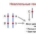 Interaction of non-allelic genes: types and forms The types of interaction of allelic genes include
 Presentation “Rules of conduct in the library