X-ray radiation properties and applications. The effect of X-rays on humans
X-RAY RADIATION
x-ray radiation occupies the region of the electromagnetic spectrum between gamma and ultraviolet radiation and is electromagnetic radiation with a wavelength of 10 -14 to 10 -7 m. X-ray radiation with a wavelength of 5 x 10 -12 to 2.5 x 10 -10 is used in medicine m, that is, 0.05 - 2.5 angstrom, and actually for X-ray diagnostics - 0.1 angstrom. Radiation is a stream of quanta (photons) propagating in a straight line at the speed of light (300,000 km/s). These quanta have no electric charge. The mass of a quantum is an insignificant part of the atomic mass unit.
Quantum energy measured in Joules (J), but in practice they often use an off-system unit "electron volt" (eV) . One electron volt is the energy that one electron acquires when it passes through a potential difference of 1 volt in an electric field. 1 eV \u003d 1.6 10 ~ 19 J. Derivatives are a kiloelectron volt (keV), equal to a thousand eV, and a megaelectron volt (MeV), equal to a million eV.
X-rays are obtained using X-ray tubes, linear accelerators and betatrons. In an X-ray tube, the potential difference between the cathode and the target anode (tens of kilovolts) accelerates the electrons bombarding the anode. X-ray radiation arises when fast electrons decelerate in the electric field of atoms of the anode substance (bremsstrahlung) or when rearranging the inner shells of atoms (characteristic radiation) . Characteristic X-rays has a discrete character and occurs when the electrons of the atoms of the anode substance pass from one energy level to another under the influence of external electrons or radiation quanta. Bremsstrahlung X-ray has a continuous spectrum depending on the anode voltage on the x-ray tube. When braking in the anode material, electrons most of their energy is spent on heating the anode (99%) and only a small fraction (1%) is converted into X-ray energy. In X-ray diagnostics, bremsstrahlung is most often used.
Basic properties x-rays characteristic of all electromagnetic radiation, but there are some features. X-rays have the following properties:
- invisibility - sensitive cells of the human retina do not react to x-rays, since their wavelength is thousands of times smaller than that of visible light;
- rectilinear propagation - rays are refracted, polarized (propagated in a certain plane) and diffracted, like visible light. The refractive index differs very little from unity;
- penetrating power - penetrate without significant absorption through significant layers of a substance that is opaque to visible light. The shorter the wavelength, the greater the penetrating power of X-rays;
- absorbency - have the ability to be absorbed by the tissues of the body, this is the basis of all x-ray diagnostics. The ability to absorb depends on the specific gravity of the tissues (the more, the greater the absorption); on the thickness of the object; on the hardness of the radiation;
- photographic action - decompose silver halide compounds, including those found in photographic emulsions, which makes it possible to obtain x-rays;
- luminescent action - cause the luminescence of a number of chemical compounds (phosphors), this is the basis of the X-ray transmission technique. The intensity of the glow depends on the structure of the fluorescent substance, its amount and distance from the source of x-rays. Phosphors are used not only to obtain an image of the objects under study on a fluoroscopic screen, but also in radiography, where they make it possible to increase the radiation exposure to a radiographic film in a cassette due to the use of intensifying screens, the surface layer of which is made of fluorescent substances;
- ionization action - have the ability to cause the decay of neutral atoms into positively and negatively charged particles, dosimetry is based on this. The effect of ionization of any medium is the formation of positive and negative ions in it, as well as free electrons from neutral atoms and molecules of matter. Ionization of air in the X-ray room during the operation of the X-ray tube leads to an increase in electrical conductivity air, strengthening static electric charges on cabinet items. In order to eliminate such an undesirable influence of them in X-ray rooms, forced supply and exhaust ventilation is provided;
- biological action - have an impact on biological objects, in most cases this impact is harmful;
- inverse square law - for a point source of X-ray radiation, the intensity decreases in proportion to the square of the distance to the source.
The discovery and merit in the study of the basic properties of X-rays rightfully belongs to the German scientist Wilhelm Conrad Roentgen. The amazing properties of X-rays discovered by him immediately received a huge response in the scientific world. Although then, back in 1895, the scientist could hardly imagine what benefit, and sometimes harm, X-rays can bring.
Let's find out in this article how this type of radiation affects human health.
What is x-ray radiation

The first question that interested the researcher was what is X-ray radiation? A number of experiments made it possible to verify that this is electromagnetic radiation with a wavelength of 10 -8 cm, which occupies an intermediate position between ultraviolet and gamma radiation.
Application of X-rays
All these aspects of the destructive effects of the mysterious X-rays do not at all exclude surprisingly extensive aspects of their application. Where is X-rays used?
- Study of the structure of molecules and crystals.
- X-ray flaw detection (in industry, detection of defects in products).
- Methods of medical research and therapy.
The most important applications of X-rays have become possible due to the very short wavelengths of the entire range of these waves and their unique properties.
Since we are interested in the effect of X-ray radiation on people who encounter it only during medical examination or treatment, then further we will consider only this area of \u200b\u200bapplication of X-rays.
The use of x-rays in medicine
 Despite the special significance of his discovery, Roentgen did not take out a patent for its use, making it an invaluable gift for all mankind. Already in the First World War, X-ray units began to be used, which made it possible to quickly and accurately diagnose the wounded. Now we can distinguish two main areas of application of x-rays in medicine:
- X-ray diagnostics;
- x-ray therapy.
X-ray diagnostics
X-ray diagnostics is used in various options:

Let's take a look at the difference between these methods.

All of these diagnostic methods are based on the ability of x-rays to illuminate film and on their different permeability to tissues and the bone skeleton.
X-ray therapy
The ability of X-rays to have a biological effect on tissues is used in medicine for the treatment of tumors. The ionizing effect of this radiation is most actively manifested in the effect on rapidly dividing cells, which are the cells of malignant tumors.
However, you should also be aware of the side effects that inevitably accompany radiotherapy. The fact is that cells of the hematopoietic, endocrine, and immune systems are also rapidly dividing. Negative impact on them gives rise to signs of radiation sickness.
The effect of X-ray radiation on humans
Shortly after the remarkable discovery of X-rays, it was discovered that X-rays had an effect on humans.

These data were obtained in experiments on experimental animals, however, geneticists suggest that similar effects may apply to the human body.
The study of the effects of X-ray exposure has led to the development of international standards for acceptable radiation doses.
Doses of x-ray radiation in x-ray diagnostics
After visiting the X-ray room, many patients are worried - how will the received dose of radiation affect their health?

The dose of general irradiation of the body depends on the nature of the procedure. For convenience, we will compare the received dose with natural exposure, which accompanies a person throughout his life.
- X-ray: chest - the received dose of radiation is equivalent to 10 days of background exposure; upper stomach and small intestine - 3 years.
- Computed tomography of the abdominal cavity and pelvis, as well as the whole body - 3 years.
- Mammography - 3 months.
- Radiography of the extremities is practically harmless.
- With regard to dental x-rays, the radiation dose is minimal, since the patient is exposed to a narrow beam of x-rays with a short radiation duration.
These radiation doses meet acceptable standards, but if the patient feels anxious before the X-ray, he has the right to ask for a special protective apron.
Exposure of X-rays to pregnant women
Each person has to undergo X-ray examination repeatedly. But there is a rule - this diagnostic method cannot be prescribed to pregnant women. The developing embryo is extremely vulnerable. X-rays can cause chromosome abnormalities and, as a result, the birth of children with malformations. The most vulnerable in this regard is the gestational age of up to 16 weeks. Moreover, the most dangerous for the future baby is an x-ray of the spine, pelvic and abdominal regions.
 Knowing about the detrimental effect of x-rays on pregnancy, doctors avoid using it in every possible way during this crucial period in a woman's life.
However, there are side sources of X-rays:
- electron microscopes;
- color TV kinescopes, etc.
Expectant mothers should be aware of the danger posed by them.
For nursing mothers, radiodiagnosis is not dangerous.
What to do after an x-ray
To avoid even the minimal effects of X-ray exposure, some simple steps can be taken:
- after an x-ray, drink a glass of milk - it removes small doses of radiation;
- very handy taking a glass of dry wine or grape juice;
- some time after the procedure, it is useful to increase the proportion of foods with a high content of iodine (seafood).
But, no medical procedures or special measures are required to remove radiation after an x-ray!
Despite the undoubtedly serious consequences of exposure to X-rays, one should not overestimate their danger during medical examinations - they are carried out only in certain areas of the body and very quickly. The benefits of them many times exceed the risk of this procedure for the human body.
Radiology is a section of radiology that studies the effects of X-ray radiation on the body of animals and humans arising from this disease, their treatment and prevention, as well as methods for diagnosing various pathologies using X-rays (X-ray diagnostics). A typical X-ray diagnostic apparatus includes a power supply (transformers), a high-voltage rectifier that converts alternating current mains into a permanent, control panel, tripod and x-ray tube.
X-rays are a type of electromagnetic oscillations that are formed in an X-ray tube during a sharp deceleration of accelerated electrons at the moment of their collision with the atoms of the anode substance. At present, the point of view is generally accepted that X-rays, by their physical nature, are one of the types of radiant energy, the spectrum of which also includes radio waves, infrared rays, visible light, ultraviolet rays and gamma rays of radioactive elements. X-ray radiation can be characterized as a collection of its smallest particles - quanta or photons.
Rice. 1 - mobile x-ray machine:
A - x-ray tube;
B - power supply;
B - adjustable tripod.
 Rice. 2 - X-ray machine control panel (mechanical - on the left and electronic - on the right):
 A - panel for adjusting exposure and hardness;
B - feed button high voltage.
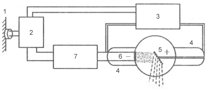 Rice. 3 is a block diagram of a typical x-ray machine
 1 - network;
2 - autotransformer;
3 - step-up transformer;
4 - x-ray tube;
5 - anode;
6 - cathode;
7 - step-down transformer.
Mechanism of X-ray generation
X-rays are formed at the moment of collision of a stream of accelerated electrons with the anode material. When electrons interact with a target, 99% of their kinetic energy is converted into thermal energy and only 1% into X-rays.
An X-ray tube consists of a glass container in which 2 electrodes are soldered: a cathode and an anode. Air is pumped out of the glass cylinder: the movement of electrons from the cathode to the anode is possible only under conditions of relative vacuum (10 -7 -10 -8 mm Hg). On the cathode there is a filament, which is a tightly twisted tungsten filament. When applying electric current electron emission occurs on the filament, in which electrons are separated from the spiral and form an electron cloud near the cathode. This cloud is concentrated at the focusing cup of the cathode, which sets the direction of electron movement. Cup - a small depression in the cathode. The anode, in turn, contains a tungsten metal plate on which the electrons are focused - this is the site of the formation of x-rays.
Rice. 4 - X-ray tube device: A - cathode; 
B - anode;
B - tungsten filament;
G - focusing cup of the cathode;
D - stream of accelerated electrons;
E - tungsten target;
G - glass flask;
З - a window from beryllium;
And - formed x-rays;
K - aluminum filter.
2 transformers are connected to the electron tube: step-down and step-up. A step-down transformer heats the tungsten coil with a low voltage (5-15 volts), resulting in electron emission. A step-up, or high-voltage, transformer goes directly to the cathode and anode, which are supplied with a voltage of 20–140 kilovolts. Both transformers are placed in the high-voltage block of the X-ray machine, which is filled with transformer oil, which provides cooling of the transformers and their reliable insulation.
After an electron cloud has formed with the help of a step-down transformer, the step-up transformer is turned on, and high-voltage voltage is applied to both poles of the electrical circuit: a positive pulse to the anode, and a negative pulse to the cathode. Negatively charged electrons are repelled from a negatively charged cathode and tend to a positively charged anode - due to such a potential difference, a high speed of movement is achieved - 100 thousand km / s. At this speed, electrons bombard the tungsten plate of the anode, shorting out electrical circuit, resulting in X-rays and thermal energy.
X-ray radiation is subdivided into bremsstrahlung and characteristic. Bremsstrahlung occurs due to a sharp slowdown in the speed of electrons emitted by a tungsten spiral. Characteristic radiation occurs at the moment of restructuring electron shells atoms. Both of these types are formed in an X-ray tube at the moment of collision of accelerated electrons with atoms of the anode material. The emission spectrum of an X-ray tube is a superposition of bremsstrahlung and characteristic X-rays.
 Rice. 5 - the principle of the formation of bremsstrahlung X-rays.
 Rice. 6 - the principle of formation of the characteristic x-rays.
Basic properties of X-rays
- X-rays are invisible to visual perception.
- X-ray radiation has a great penetrating power through the organs and tissues of a living organism, as well as dense structures of inanimate nature, which do not transmit visible light rays.
- X-rays cause certain chemical compounds to glow, called fluorescence.
- Zinc and cadmium sulfides fluoresce yellow-green,
- Crystals of calcium tungstate - violet-blue.
Scale of electromagnetic oscillations
X-rays have certain length waves and vibration frequency. Wavelength (λ) and oscillation frequency (ν) are related by the relationship: λ ν = c, where c is the speed of light, rounded to 300,000 km per second. The energy of X-rays is determined by the formula E = h ν, where h is Planck's constant, a universal constant equal to 6.626 10 -34 J⋅s. The wavelength of the rays (λ) is related to their energy (E) by the relation: λ = 12.4 / E.
X-ray radiation differs from other types of electromagnetic oscillations in wavelength (see table) and quantum energy. The shorter the wavelength, the higher its frequency, energy and penetrating power. The X-ray wavelength is in the range
. By changing the wavelength of X-ray radiation, it is possible to control its penetrating power. X-rays have a very short wavelength, but a high frequency of oscillation, so they are invisible to the human eye. Due to their enormous energy, quanta have a high penetrating power, which is one of the main properties that ensure the use of X-rays in medicine and other sciences.X-ray characteristics
Intensity- quantitative characteristic of x-ray radiation, which is expressed by the number of rays emitted by the tube per unit time. The intensity of X-rays is measured in milliamps. Comparing it with the intensity of visible light from a conventional incandescent lamp, we can draw an analogy: for example, a 20-watt lamp will shine with one intensity, or power, and a 200-watt lamp will shine with another, while the quality of the light itself (its spectrum) is the same . The intensity of X-ray radiation is, in fact, its quantity. Each electron creates one or more radiation quanta on the anode, therefore, the amount of X-rays during exposure of the object is regulated by changing the number of electrons tending to the anode and the number of interactions of electrons with atoms of the tungsten target, which can be done in two ways:
- By changing the degree of incandescence of the cathode spiral using a step-down transformer (the number of electrons generated during emission will depend on how hot the tungsten spiral is, and the number of radiation quanta will depend on the number of electrons);
- By changing the value of the high voltage supplied by the step-up transformer to the poles of the tube - the cathode and the anode (the higher the voltage is applied to the poles of the tube, the more kinetic energy the electrons receive, which, due to their energy, can interact with several atoms of the anode substance in turn - see Fig. rice. 5; low energy electrons can enter into lesser number interactions).
The X-ray intensity (anode current) multiplied by the exposure (tube time) corresponds to the X-ray exposure, which is measured in mAs (milliamps per second). Exposure is a parameter that, like intensity, characterizes the amount of rays emitted by an x-ray tube. The only difference is that the exposure also takes into account the operating time of the tube (for example, if the tube works for 0.01 sec, then the number of rays will be one, and if 0.02 sec, then the number of rays will be different - twice more). The radiation exposure is set by the radiologist on the control panel of the X-ray machine, depending on the type of examination, the size of the object under study and the diagnostic task.
Rigidity- qualitative characteristic of x-ray radiation. It is measured by the high voltage on the tube - in kilovolts. Determines the penetrating power of x-rays. It is regulated by the high voltage supplied to the X-ray tube by a step-up transformer. The higher the potential difference is created on the electrodes of the tube, the more force the electrons repel from the cathode and rush to the anode, and the stronger their collision with the anode. The stronger their collision, the shorter the wavelength of the resulting X-ray radiation and the higher the penetrating power of this wave (or the hardness of the radiation, which, like the intensity, is regulated on the control panel by the voltage parameter on the tube - kilovoltage).
λ - wavelength;  Rice. 7 - Dependence of the wavelength on the energy of the wave:
E - wave energy  Rice. 8 - The ratio of the voltage on the X-ray tube and the wavelength of the resulting X-ray radiation:
Classification of x-ray tubes
- By appointment
- Diagnostic
- Therapeutic
- For structural analysis
- For transillumination
- By design
- By focus
- Single-focus (one spiral on the cathode, and one focal spot on the anode)
- Bifocal (two spirals of different sizes on the cathode, and two focal spots on the anode)
- By type of anode
- Stationary (fixed)
- Rotating
X-rays are used not only for radiodiagnostic purposes, but also for therapeutic purposes. As noted above, the ability of X-ray radiation to suppress the growth of tumor cells makes it possible to use it in radiation therapy of oncological diseases. In addition to the medical field of application, X-ray radiation has found wide application in the engineering and technical field, materials science, crystallography, chemistry and biochemistry: for example, it is possible to identify structural defects in various products (rails, welds, etc.) using X-ray radiation. The type of such research is called defectoscopy. And at airports, railway stations and other crowded places, X-ray television introscopes are actively used to scan hand luggage and luggage for security purposes.
Depending on the type of anode, X-ray tubes differ in design. Due to the fact that 99% of the kinetic energy of the electrons is converted into thermal energy, during the operation of the tube, the anode is significantly heated - the sensitive tungsten target often burns out. The anode is cooled in modern x-ray tubes by rotating it. The rotating anode has the shape of a disk, which distributes heat evenly over its entire surface, preventing local overheating of the tungsten target.
The design of X-ray tubes also differs in focus. Focal spot - the section of the anode on which the working X-ray beam is generated. It is subdivided into the real focal spot and the effective focal spot ( rice. 12). Due to the angle of the anode, the effective focal spot is smaller than the real one. Different focal spot sizes are used depending on the size of the image area. How more area image, the wider the focal spot must be to cover the entire area of the image. However, a smaller focal spot produces better image clarity. Therefore, when producing small images, a short filament is used and the electrons are directed to a small area of the anode target, creating a smaller focal spot.
 Rice. 9 - x-ray tube with a stationary anode.
 Rice. 10 - X-ray tube with a rotating anode.
 Rice. 11 - X-ray tube device with a rotating anode.
 Rice. 12 is a diagram of the formation of a real and effective focal spot.
FEDERAL AGENCY FOR EDUCATION OF THE RUSSIAN FEDERATION
STATE EDUCATIONAL INSTITUTION
HIGHER PROFESSIONAL EDUCATION
MOSCOW STATE INSTITUTE OF STEEL AND ALLOYS
(UNIVERSITY OF TECHNOLOGY)
NOVOTROITSKY BRANCH
Department of OEND
COURSE WORK
Discipline: Physics
Topic: X-RAY
Student: Nedorezova N.A.
Group: EiU-2004-25, No. З.К.: 04Н036
Checked by: Ozhegova S.M.
Introduction
Chapter 1
1.1 Biography of Roentgen Wilhelm Conrad
1.2 Discovery of X-rays
Chapter 2
2.1 X-ray sources
2.2 Properties of X-rays
2.3 Registration of X-rays
2.4 Use of X-rays
Chapter 3
3.1 Imperfection analysis crystal structure
3.2 Spectrum analysis
Conclusion
List of sources used
Applications
Introduction
A rare person has not gone through an x-ray room. Pictures taken in x-rays are familiar to everyone. In 1995, this discovery was 100 years old. It is hard to imagine what great interest it aroused a century ago. In the hands of a man turned out to be an apparatus with which it was possible to see the invisible.
This invisible radiation capable of penetrating, albeit to varying degrees, into all substances, which is electromagnetic radiation with a wavelength of about 10 -8 cm, was called X-ray radiation, in honor of Wilhelm Roentgen who discovered it.
Like visible light, X-rays cause blackening of photographic film. This property is of great importance for medicine, industry and scientific research. Passing through the object under study and then falling on the film, X-ray radiation depicts its internal structure on it. Since the penetrating power of X-rays is different for different materials, parts of the object that are less transparent to it give brighter areas in the photograph than those through which the radiation penetrates well. Thus, bone tissues are less transparent to x-rays than the tissues that make up the skin and internal organs. Therefore, on the radiograph, the bones will be indicated as lighter areas and the fracture site, which is less transparent for radiation, can be quite easily detected. X-ray imaging is also used in dentistry to detect caries and abscesses in the roots of teeth, as well as in industry to detect cracks in castings, plastics and rubbers, in chemistry to analyze compounds, and in physics to study the structure of crystals.
Roentgen's discovery was followed by experiments by other researchers who discovered many new properties and possibilities for using this radiation. A major contribution was made by M. Laue, W. Friedrich, and P. Knipping, who in 1912 demonstrated the diffraction of X-rays as they pass through a crystal; W. Coolidge, who in 1913 invented a high-vacuum x-ray tube with a heated cathode; G. Moseley, who established in 1913 the relationship between the wavelength of radiation and the atomic number of an element; G. and L. Braggi, who received the Nobel Prize in 1915 for developing the fundamentals of X-ray diffraction analysis.
The purpose of this course work is to study the phenomenon of x-ray radiation, the history of discovery, properties and identify the scope of its application.
Chapter 1
1.1 Biography of Roentgen Wilhelm Conrad
Wilhelm Conrad Roentgen was born on March 17, 1845 in the border region of Germany with Holland, in the city of Lenepe. He received his technical education in Zurich at the same Higher Technical School (Polytechnic) where Einstein later studied. Passion for physics forced him after leaving school in 1866 to continue physical education.
In 1868 he defended his dissertation for the degree of Doctor of Philosophy, he worked as an assistant at the Department of Physics, first in Zurich, then in Giessen, and then in Strasbourg (1874-1879) with Kundt. Here Roentgen went through a good experimental school and became a first-class experimenter. Roentgen performed part of the important research with his student, one of the founders of Soviet physics, A.F. Ioffe.
Scientific research relates to electromagnetism, crystal physics, optics, molecular physics.
In 1895, he discovered radiation with a wavelength shorter than the wavelength of ultraviolet rays (X-rays), later called x-rays, and investigated their properties: the ability to reflect, absorb, ionize air, etc. He proposed the correct design of the tube for obtaining X-rays - an inclined platinum anticathode and a concave cathode: he was the first to take photographs using X-rays. He discovered in 1885 the magnetic field of a dielectric moving in an electric field (the so-called "roentgen current"). His experience clearly showed that a magnetic field is created by moving charges, and was important for the creation of X. Lorentz's electronic theory. A significant number of Roentgen's works are devoted to the study properties of liquids, gases, crystals, electromagnetic phenomena, discovered the relationship between electrical and optical phenomena in crystals. For the discovery of the rays that bear his name, Roentgen in 1901 was the first among physicists to be awarded the Nobel Prize.
From 1900 until the last days of his life (he died on February 10, 1923) he worked at the University of Munich.
1.2 Discovery of X-rays
End of the 19th century was marked by increased interest in the phenomena of the passage of electricity through gases. Even Faraday seriously studied these phenomena, described various forms of discharge, discovered a dark space in a luminous column of rarefied gas. Faraday dark space separates the bluish, cathode glow from the pinkish, anode glow.
A further increase in the rarefaction of the gas significantly changes the nature of the glow. The mathematician Plücker (1801-1868) discovered in 1859, at sufficiently strong rarefaction, a weakly bluish beam of rays emanating from the cathode, reaching the anode and causing the glass of the tube to glow. Plücker's student Gittorf (1824-1914) in 1869 continued his teacher's research and showed that a distinct shadow appears on the fluorescent surface of the tube if a solid body is placed between the cathode and this surface.
Goldstein (1850-1931), studying the properties of rays, called them cathode rays (1876). Three years later, William Crookes (1832-1919) proved the material nature of cathode rays and called them "radiant matter" - a substance in a special fourth state. His evidence was convincing and clear. Experiments with the "Crookes tube" were demonstrated later in all physical classrooms . The deflection of the cathode beam by a magnetic field in a Crookes tube has become a classic school demonstration.
However, experiments on the electrical deflection of cathode rays were not so convincing. Hertz did not detect such a deviation and came to the conclusion that the cathode ray is an oscillatory process in the ether. Hertz's student F. Lenard, experimenting with cathode rays, showed in 1893 that they pass through a window covered with aluminum foil and cause a glow in the space behind the window. The phenomenon of the passage of cathode rays through thin metal bodies Hertz devoted his latest article, published in 1892. It began with the words:
“Cathode rays differ from light in a significant way in terms of their ability to penetrate solids.” Describing the results of experiments on the passage of cathode rays through gold, silver, platinum, aluminum, etc. leaves, Hertz notes that he did not observe any special differences in the phenomena The rays do not pass through the leaves in a straight line, but are scattered by diffraction.The nature of the cathode rays was still unclear.
It was with such tubes of Crookes, Lenard and others that the Würzburg professor Wilhelm Conrad Roentgen experimented at the end of 1895. Once, after the end of the experiment, he closed the tube with a black cardboard cover, turned off the light, but did not turn off the inductor that fed the tube, he noticed the glow of the screen from barium cyanogen located near the tube. Struck by this circumstance, Roentgen began to experiment with the screen. In his first report "On a new kind of rays", dated December 28, 1895, he wrote about these first experiments: "A piece of paper coated with barium platinum-cyanide, when approaching a tube, closed with a cover of thin black cardboard that fits snugly enough to it, with each discharge it flashes with a bright light: it begins to fluoresce. Fluorescence is visible with sufficient darkening and does not depend on whether we bring the paper with the side coated with barium synerogen or not coated with barium synerogen. The fluorescence is noticeable even at a distance of two meters from the tube.”
Careful examination showed Roentgen "that black cardboard, transparent neither to the visible and ultraviolet rays of the sun, nor to the rays of an electric arc, is penetrated by some kind of fluorescent agent." Roentgen investigated the penetrating power of this "agent", which he called for brevity "X-rays", for various substances. He found that the rays freely pass through paper, wood, ebonite, thin layers of metal, but are strongly delayed by lead.
He then describes the sensational experience:
“If you hold your hand between the discharge tube and the screen, you can see the dark shadows of the bones in the faint outlines of the shadow of the hand itself.” This was the first x-ray examination of the human body. Roentgen also received the first x-rays by attaching them to his hand.
These shots made a huge impression; the discovery had not yet been completed, and X-ray diagnostics had already begun its journey. “My laboratory was flooded with doctors bringing in patients who suspected they had needles in various parts of their bodies,” wrote the English physicist Schuster.
Already after the first experiments, Roentgen firmly established that X-rays differ from cathode ones, they do not carry a charge and are not deflected by a magnetic field, but they are excited by cathode rays. "X-rays are not identical with cathode rays, but they are excited by them in the glass walls of the discharge tube ”, wrote Roentgen.
He also established that they are excited not only in glass, but also in metals.
Mentioning the Hertz-Lenard hypothesis that cathode rays “are a phenomenon occurring in the ether,” Roentgen points out that “we can say something similar about our rays.” However, he failed to find wave properties rays, they "behave differently than hitherto known ultraviolet, visible, infrared rays." In their chemical and luminescent actions, they, according to Roentgen, are similar to ultraviolet rays. In the first message, he expressed the assumption left later that they can be longitudinal waves on the air.
Roentgen's discovery aroused great interest in the scientific world. His experiments were repeated in almost all laboratories in the world. In Moscow they were repeated by P.N. Lebedev. In St. Petersburg, the inventor of radio A.S. Popov experimented with X-rays, demonstrated them on public lectures while taking different radiographs. In Cambridge D.D. Thomson immediately applied the ionizing effect of X-rays to study the passage of electricity through gases. His research led to the discovery of the electron.
Chapter 2
X-ray radiation - electromagnetic ionizing radiation, occupying the spectral region between gamma and ultraviolet radiation within wavelengths from 10 -4 to 10 3 (from 10 -12 to 10 -5 cm).R. l. with wavelength λ< 2 условно называются жёсткими, с λ >2 - soft.
2.1 X-ray sources
The most common source of X-rays is the X-ray tube.
X-ray tubes are used in X-ray structural analysis
The main characteristics of X-ray tubes are the maximum permissible accelerating voltage (1-500 kV), electronic current (0.01 mA - 1A), specific power dissipated by the anode (10-10 4 W / mm 2), total power consumption (0.002 W - 60 kW) and focus sizes (1 µm - 10 mm). The efficiency of the x-ray tube is 0.1-3%.
Some radioactive isotopes can also serve as sources of X-rays.
Synchrotrons and electron storage rings with energies of several GeV can serve as sources of soft X-rays with λ on the order of tens and hundreds. In intensity, the X-ray radiation of synchrotrons exceeds the radiation of an X-ray tube in the specified region of the spectrum by 2-3 orders of magnitude.
Natural sources of X-rays - the Sun and other space objects.
2.2 Properties of X-rays
Depending on the mechanism of origin of X-rays, their spectra can be continuous (bremsstrahlung) or line (characteristic). A continuous X-ray spectrum is emitted by fast charged particles as a result of their deceleration when interacting with target atoms; this spectrum reaches a significant intensity only when the target is bombarded with electrons. The intensity of bremsstrahlung X-rays is distributed over all frequencies up to the high-frequency boundary 0 , at which the photon energy h 0 (h is Planck's constant
Line radiation occurs after the ionization of an atom with the ejection of an electron from one of its inner shells. Such ionization can be the result of an atom colliding with a fast particle, such as an electron (primary x-rays), or the absorption of a photon by an atom (fluorescent x-rays). The ionized atom finds itself in the initial quantum state on one of high levels energy and after 10 -16 -10 -15 seconds goes into the final state with less energy. In this case, an atom can emit an excess of energy in the form of a photon of a certain frequency. The frequencies of the lines of the spectrum of such radiation are characteristic of the atoms of each element, therefore the line X-ray spectrum is called characteristic. The dependence of the line frequency of this spectrum on the atomic number Z is determined by the Moseley law.
Moseley's law, the law relating the frequency of the spectral lines of the characteristic X-ray emission of a chemical element with its serial number. G. Moseley experimentally installed
where R is the Rydberg constant
Moseley's law was irrefutable proof of the correct placement of elements in the periodic table of elements
In accordance with Moseley's law, X-ray characteristic spectra do not exhibit the periodic patterns inherent in optical spectra. This indicates that the inner electron shells of atoms of all elements that appear in the characteristic X-ray spectra have a similar structure.
Later experiments revealed some deviations from the linear dependence for the transition groups of elements, associated with a change in the order of filling of the outer electron shells, as well as for heavy atoms, resulting from relativistic effects (conditionally explained by the fact that the speeds of the inner ones are comparable to the speed of light).
Depending on a number of factors - on the number of nucleons in the nucleus (isotonic shift), the state of the outer electron shells (chemical shift), etc. - the position of the spectral lines on the Moseley diagram may change somewhat. The study of these shifts allows one to obtain detailed information about the atom.
Bremsstrahlung X-rays emitted by very thin targets are completely polarized near 0; as 0 decreases, the degree of polarization decreases. Characteristic radiation, as a rule, is not polarized.
When X-rays interact with matter, the photoelectric effect can occur.
When X-rays pass through a layer of substance with thickness x, their initial intensity I 0 decreases to the value I = I 0 e - μ x where μ is the attenuation coefficient. The attenuation of I occurs due to two processes: the absorption of X-ray photons by matter and the change in their direction upon scattering. In the long-wavelength region of the spectrum, the absorption of X-rays predominates, in the short-wavelength region, their scattering. The degree of absorption increases rapidly with increasing Z and λ. For example, hard X-rays freely penetrate through a layer of air ~ 10 cm; an aluminum plate 3 cm thick attenuates X-rays with λ = 0.027 by half; soft x-rays are significantly absorbed in air and their use and study is possible only in a vacuum or in a weakly absorbing gas (for example, He). When X-rays are absorbed, the atoms of a substance are ionized.
The effect of X-rays on living organisms can be beneficial or harmful, depending on the ionization they cause in the tissues. Since the absorption of X-rays depends on λ, their intensity cannot serve as a measure of the biological effect of X-rays. X-ray measurements are used to measure the effect of X-rays on matter.
Scattering of X-rays in the region of large Z and λ occurs mainly without a change in λ and is called coherent scattering, and in the region of small Z and λ, as a rule, it increases (incoherent scattering). There are 2 types of incoherent X-ray scattering - Compton and Raman. In Compton scattering, which has the character of inelastic corpuscular scattering, a recoil electron flies out of the atomic shell due to the energy partially lost by the X-ray photon. In this case, the energy of the photon decreases and its direction changes; the change in λ depends on the scattering angle. During Raman scattering of a high-energy X-ray photon by a light atom, a small part of its energy is spent on ionization of the atom and the direction of the photon's motion changes. The change of such photons does not depend on the scattering angle.
The refractive index n for x-rays differs from 1 by a very small amount δ = 1-n ≈ 10 -6 -10 -5 . The phase velocity of X-rays in a medium is greater than the speed of light in a vacuum. The deviation of X-rays during the transition from one medium to another is very small (a few arc minutes). When X-rays fall from a vacuum onto the surface of a body at a very small angle, their total external reflection occurs.
2.3 Registration of X-rays
The human eye is not sensitive to x-rays. X-ray
rays are recorded using a special x-ray film containing an increased amount of Ag, Br. In the region λ<0,5 чувствительность этих плёнок быстро падает и может быть
искусственно повышена плотно прижатым к плёнке флуоресцирующим экраном. В
области λ>5, the sensitivity of ordinary positive film is quite high, and its grains are much smaller than the grains of X-ray film, which increases the resolution. At λ of the order of tens and hundreds, X-rays act only on the thinnest surface layer of the photographic emulsion; to increase the sensitivity of the film, it is sensitized with luminescent oils. In X-ray diagnostics and flaw detection, electrophotography is sometimes used to record X-rays.
X-rays of high intensity can be recorded using an ionization chamber
2.4 Use of X-rays
X-rays are most widely used in medicine for X-ray diagnostics.
X-ray structural analysis
X-ray microscopy
X-rays coming from space carry information about the chemical composition of cosmic bodies and about physical processes taking place in space. X-ray astronomy deals with the study of cosmic x-rays
Chapter 3
One of the main tasks of X-ray diffraction analysis is the determination of the real or phase composition of a material. The X-ray diffraction method is direct and is characterized by high reliability, rapidity and relative cheapness. The method does not require a large number substances, the analysis can be carried out without destroying the part. The areas of application of qualitative phase analysis are very diverse both for scientific research and for control in production. You can check the composition of the raw materials of metallurgical production, synthesis products, processing, the result of phase changes during thermal and chemical-thermal treatment, analyze various coatings, thin films, etc.
Each phase, having its own crystal structure, is characterized by a certain set of discrete values of interplanar distances d/n from the maximum and below, inherent only to this phase. As follows from the Wulf-Bragg equation, each value of the interplanar distance corresponds to a line on the x-ray pattern from a polycrystalline sample at a certain angle θ (at a given value of the wavelength λ). Thus, a certain system of lines (diffraction maxima) will correspond to a certain set of interplanar distances for each phase in the X-ray diffraction pattern. The relative intensity of these lines in the X-ray pattern depends primarily on the structure of the phase. Therefore, by determining the location of the lines on the X-ray image (its angle θ) and knowing the wavelength of the radiation on which the X-ray image was taken, it is possible to determine the values of the interplanar distances d/n using the Wulf-Bragg formula:
/n = λ/ (2sin θ). (one)
Having determined the set of d/n for the material under study and comparing it with the previously known d/n data for pure substances, their various compounds, it is possible to establish which phase the given material constitutes. It should be emphasized that it is the phases that are determined, and not chemical composition, but the latter can sometimes be derived if there are additional data on the elemental composition of a particular phase. The task of qualitative phase analysis is greatly facilitated if the chemical composition of the material under study is known, because then it is possible to make preliminary assumptions about the possible this case phases.
The key to phase analysis is to accurately measure d/n and line intensity. Although this is in principle easier to achieve using a diffractometer, the photomethod for qualitative analysis has some advantages, primarily in terms of sensitivity (the ability to detect the presence of a small amount of phase in the sample), as well as the simplicity of the experimental technique.
The calculation of d/n from the X-ray pattern is carried out using the Wulf-Bragg equation.
As the value of λ in this equation, λ α cf K-series is usually used:
λ α cf = (2λ α1 + λ α2) /3 (2)
Sometimes the K α1 line is used. Determining the diffraction angles θ for all X-ray lines allows you to calculate d / n according to equation (1) and separate the β-lines (if there was no filter for (β-rays).
3.1 Analysis of crystal structure imperfections
All real single-crystal and even more so polycrystalline materials contain certain structural imperfections (point defects, dislocations, various types of interfaces, micro- and macrostresses), which have a very strong influence on all structure-sensitive properties and processes.
Structural imperfections cause distortions of the crystal lattice of different nature and, as a result, different types of changes in the diffraction pattern: a change in interatomic and interplanar distances causes a shift in diffraction maxima, microstresses and dispersity of the substructure lead to a broadening of diffraction maxima, lattice microdistortions - to a change in the intensity of these maxima, the presence dislocations causes anomalous phenomena during the passage of X-rays and, consequently, local contrast inhomogeneities on X-ray topograms, etc.
As a result, X-ray diffraction analysis is one of the most informative methods for studying structural imperfections, their type and concentration, and the nature of their distribution.
The traditional direct method of X-ray diffraction, which is implemented on stationary diffractometers, due to their design features, allows quantitative determination of stresses and strains only on small samples cut from parts or objects.
Therefore, at present, there is a transition from stationary to portable small-sized X-ray diffractometers, which provide an assessment of stresses in the material of parts or objects without destruction at the stages of their manufacture and operation.
Portable X-ray diffractometers of the DRP * 1 series make it possible to control residual and effective stresses in large-sized parts, products and structures without destruction
The program in the Windows environment allows not only to determine the stresses using the "sin 2 ψ" method in real time, but also to monitor the change in the phase composition and texture. The linear coordinate detector provides simultaneous registration at diffraction angles 2θ = 43°. small-sized X-ray tubes of the "Fox" type with high luminosity and low power (5 W) ensure the radiological safety of the device, in which at a distance of 25 cm from the irradiated area, the radiation level is equal to the natural background level. Devices of the DRP series are used in determining stresses at various stages of metal forming, cutting, grinding, heat treatment, welding, surface hardening in order to optimize these technological operations. Control over the drop in the level of induced residual compressive stresses in especially critical products and structures during their operation makes it possible to take the product out of service before its destruction, preventing possible accidents and catastrophes.
3.2 Spectrum analysis
Along with the determination of the atomic crystal structure and phase composition of the material for its complete characteristics it is mandatory to define chemical composition.
Increasingly, various so-called instrumental methods of spectral analysis are used in practice for these purposes. Each of them has its own advantages and applications.
One of the important requirements in many cases is that the method used ensures the safety of the analyzed object; It is these methods of analysis that are discussed in this section. The next criterion according to which the methods of analysis described in this section were chosen is their locality.
The method of fluorescence X-ray spectral analysis is based on the penetration of rather hard X-ray radiation (from an X-ray tube) into the analyzed object, penetrating into a layer with a thickness of the order of several micrometers. The characteristic X-ray radiation arising in this case in the object makes it possible to obtain averaged data on its chemical composition.
To determine the elemental composition of a substance, one can use the analysis of the characteristic X-ray spectrum of a sample placed on the anode of an X-ray tube and subjected to electron bombardment - the emission method, or the analysis of the spectrum of secondary (fluorescent) X-ray radiation of a sample subjected to irradiation with hard X-rays from an X-ray tube or other source - fluorescent method.
disadvantage emission method is, firstly, the need to place the sample on the anode of the X-ray tube, followed by pumping out with vacuum pumps; obviously, this method is unsuitable for fusible and volatile substances. The second drawback is related to the fact that even refractory objects are damaged by electron bombardment. The fluorescent method is free from these shortcomings and therefore has a much wider application. The advantage of the fluorescence method is also the absence of bremsstrahlung, which improves the sensitivity of the analysis. Comparison of the measured wavelengths with tables of spectral lines of chemical elements is the basis of a qualitative analysis, and the relative intensities of the spectral lines of different elements that form the sample substance form the basis of a quantitative analysis. From a consideration of the mechanism of excitation of characteristic X-ray radiation, it is clear that the radiations of one or another series (K or L, M, etc.) arise simultaneously, and the ratio of line intensities within the series is always constant. Therefore, the presence of this or that element is established not by individual lines, but by a series of lines as a whole (except for the weakest ones, taking into account the content of this element). For relatively light elements, the analysis of the K-series lines is used, for heavy elements, the L-series lines; in different conditions(depending on the instrumentation used and the elements being analyzed) different regions of the characteristic spectrum may be most convenient.
The main features of X-ray spectral analysis are as follows.
Simplicity of X-ray characteristic spectra even for heavy elements (compared to optical spectra), which simplifies the analysis (a small number of lines; similarity in their mutual arrangement; with an increase in the serial number, a regular shift of the spectrum to the short-wavelength region occurs; the comparative simplicity of quantitative analysis).
Independence of wavelengths from the state of atoms of the analyzed element (free or in a chemical compound). This is due to the fact that the occurrence of characteristic X-ray radiation is associated with the excitation of internal electronic levels, which in most cases practically do not change with the degree of ionization of atoms.
The possibility of separation in the analysis of rare earth and some other elements that have small differences in the spectra in the optical range due to similarity electronic structure outer shells and differ very little in their chemical properties.
X-ray fluorescence spectroscopy is "non-destructive", so it has an advantage over conventional optical spectroscopy when analyzing thin samples - thin metal sheet, foil, etc.
X-ray fluorescence spectrometers, among them multichannel spectrometers or quantometers, providing express quantitative analysis of elements (from Na or Mg to U) with an error of less than 1% of the determined value, a sensitivity threshold of 10 -3 ... 10 -4% .
x-ray beam
Methods for determining the spectral composition of x-rays
Spectrometers are divided into two types: crystal-diffraction and crystalless.
Decomposition of X-rays into a spectrum using natural grating- crystal - essentially similar to obtaining a spectrum of ordinary light rays using an artificial diffraction grating in the form of periodic strokes on glass. The condition for the formation of a diffraction maximum can be written as the condition of "reflection" from a system of parallel atomic planes separated by a distance d hkl .
When conducting a qualitative analysis, one can judge the presence of an element in a sample by one line - usually the most intense line of the spectral series suitable for a given analyzer crystal. The resolution of crystal diffraction spectrometers is sufficient to separate the characteristic lines even of elements adjacent in position in the periodic table. However, it is also necessary to take into account the imposition of different lines of different elements, as well as the imposition of reflections of different orders. This circumstance should be taken into account when choosing analytical lines. At the same time, it is necessary to use the possibilities of improving the resolution of the device.
Conclusion
Thus, x-rays are invisible electromagnetic radiation with a wavelength of 10 5 - 10 2 nm. X-rays can penetrate some materials that are opaque to visible light. They are emitted during the deceleration of fast electrons in matter (continuous spectrum) and during transitions of electrons from the outer electron shells of the atom to the inner ones (linear spectrum). Sources of x-rays are: x-ray tube, some radioactive isotopes, accelerators and electron accumulators ( synchrotron radiation). Receivers - film, luminescent screens, nuclear radiation detectors. X-rays are used in X-ray diffraction analysis, medicine, flaw detection, X-ray spectral analysis, etc.
Having considered positive sides the discovery of V. Roentgen, it is necessary to note its harmful biological effect. It turned out that X-rays can cause something like a severe sunburn (erythema), accompanied, however, by deeper and more permanent damage to the skin. Appearing ulcers often turn into cancer. In many cases, fingers or hands had to be amputated. There were also deaths.
It has been found that skin damage can be avoided by reducing exposure time and dose, using shielding (eg lead) and remote controls. But gradually other, more long-term effects of X-ray exposure were revealed, which were then confirmed and studied in experimental animals. Effects due to X-rays and other ionizing radiations (such as gamma rays emitted by radioactive materials) include:
) temporary changes in the composition of the blood after a relatively small excess exposure;
) irreversible changes in the composition of the blood (hemolytic anemia) after prolonged excessive exposure;
) an increase in the incidence of cancer (including leukemia);
) faster aging and early death;
) the occurrence of cataracts.
The biological impact of X-rays on the human body is determined by the level of radiation dose, as well as by which particular organ of the body was exposed to radiation.
The accumulation of knowledge about the effects of X-ray radiation on the human body has led to the development of national and international standards for permissible radiation doses, published in various reference publications.
To avoid harmful effects X-ray radiation control methods are used:
) availability of adequate equipment,
) monitoring compliance with safety regulations,
) correct use of the equipment.
List of sources used
1) Blokhin M.A., Physics of X-rays, 2nd ed., M., 1957;
) Blokhin M.A., Methods of X-ray spectral studies, M., 1959;
) X-rays. Sat. ed. M.A. Blokhin, trans. with him. and English, M., 1960;
) Kharaja F., General course X-ray engineering, 3rd ed., M. - L., 1966;
) Mirkin L.I., Handbook of X-ray diffraction analysis of polycrystals, M., 1961;
) Weinstein E.E., Kakhana M.M., Reference tables on X-ray spectroscopy, M., 1953.
) X-ray and electron-optical analysis. Gorelik S.S., Skakov Yu.A., Rastorguev L.N.: Proc. Allowance for universities. - 4th ed. Add. And a reworker. - M.: "MISiS", 2002. - 360 p.
Applications
Appendix 1
General view of X-ray tubes

Annex 2
Scheme of X-ray tube for structural analysis

Scheme of an X-ray tube for structural analysis: 1 - metal anode glass (usually grounded); 2 - windows made of beryllium for x-ray output; 3 - thermionic cathode; 4 - glass bulb, isolating the anode part of the tube from the cathode; 5 - cathode terminals, to which the filament voltage is applied, as well as high (relative to the anode) voltage; 6 - electrostatic system for focusing electrons; 7 - anode (anticathode); 8 - branch pipes for input and output of running water cooling the anode glass.
Annex 3
Moseley diagram

Moseley diagram for K-, L- and M-series of characteristic X-rays. The abscissa shows the serial number of the element Z, the ordinate - ( with is the speed of light).
Appendix 4
Ionization chamber.

Fig.1. Section of a cylindrical ionization chamber: 1 - cylindrical body of the chamber, which serves as a negative electrode; 2 - cylindrical rod serving as a positive electrode; 3 - insulators.

Rice. 2. Scheme of switching on the current ionization chamber: V - voltage on the electrodes of the chamber; G is a galvanometer that measures the ionization current.

Rice. 3. Current-voltage characteristic of the ionization chamber.

Rice. 4. Scheme of switching on the pulsed ionization chamber: C - capacitance of the collecting electrode; R is resistance.
Annex 5
Scintillation counter.

Scheme of a scintillation counter: light quanta (photons) "knock out" electrons from the photocathode; moving from dynode to dynode, the electron avalanche multiplies.
Appendix 6
Geiger-Muller counter.

Rice. 1. Scheme of a glass Geiger-Muller counter: 1 - hermetically sealed glass tube; 2 - cathode (a thin layer of copper on a stainless steel tube); 3 - output of the cathode; 4 - anode (thin stretched thread).

Rice. 2. Scheme of switching on the Geiger-Muller counter.

Rice. 3. The counting characteristic of the Geiger-Muller counter.
Annex 7
proportional counter.

Scheme of a proportional counter: a - electron drift region; b - area of gas amplification.
Annex 8
Semiconductor detectors
Semiconductor detectors; the sensitive area is highlighted by hatching; n - region of a semiconductor with electronic conductivity, p - with hole, i - with intrinsic conduction; a - silicon surface-barrier detector; b - drift germanium-lithium planar detector; c - germanium-lithium coaxial detector.
Modern medical diagnostics and treatment of certain diseases cannot be imagined without devices that use the properties of X-rays. The discovery of X-rays occurred more than 100 years ago, but even now work continues on the creation of new methods and apparatus to minimize the negative effect of radiation on the human body.
Who and how discovered X-rays
Under natural conditions, the flux of X-rays is rare and is emitted only by some radioactive isotopes. X-rays or X-rays were only discovered in 1895 by the German scientist Wilhelm Röntgen. This discovery happened by chance, during an experiment to study the behavior of light rays under conditions approaching vacuum. The experiment involved a cathode gas discharge tube with reduced pressure and a fluorescent screen, which each time began to glow at the moment when the tube began to act.
Interested in a strange effect, Roentgen conducted a series of studies showing that the resulting visible to the eye radiation is able to penetrate various obstacles: paper, wood, glass, some metals, and even through human body. Despite the lack of understanding of the very nature of what is happening, whether such a phenomenon is caused by the generation of a stream of unknown particles or waves, the following pattern was noted - radiation easily passes through the soft tissues of the body, and much harder through solid living tissues and inanimate substances.
Roentgen was not the first to study this phenomenon. AT mid-nineteenth centuries, similar possibilities were studied by the Frenchman Antoine Mason and the Englishman William Crookes. However, it was Roentgen who first invented the cathode tube and an indicator that could be used in medicine. He first published treatise, which brought him the title of the first Nobel laureate among physicists.
In 1901, a fruitful collaboration began between the three scientists, who became the founding fathers of radiology and radiology.

X-ray properties
X-rays are part of the total spectrum electromagnetic radiation. The wavelength is between gamma and ultraviolet rays. X-rays have all the usual wave properties:
- diffraction;
- refraction;
- interference;
- propagation speed (it is equal to light).
To artificially generate an X-ray flux, special devices are used - X-ray tubes. X-ray radiation arises from the contact of fast tungsten electrons with substances evaporating from a hot anode. Against the background of interaction, short-length electromagnetic waves arise, which are in the spectrum from 100 to 0.01 nm and in the energy range of 100-0.1 MeV. If the wavelength of the rays is less than 0.2 nm - this is hard radiation, if the wavelength is greater than the specified value, they are called soft x-rays.
It is significant that the kinetic energy arising from the contact of electrons and the anode substance is 99% converted into heat energy and only 1% is X-rays.
X-ray radiation - bremsstrahlung and characteristic
X-radiation is a superposition of two types of rays - bremsstrahlung and characteristic. They are generated in the handset simultaneously. Therefore, X-ray irradiation and the characteristic of each specific X-ray tube - the spectrum of its radiation, depends on these indicators, and represents their superposition.
Bremsstrahlung or continuous X-rays are the result of deceleration of electrons evaporating from a tungsten filament.
Characteristic or line X-rays are formed at the moment of rearrangement of the atoms of the substance of the anode of the X-ray tube. The wavelength of the characteristic rays directly depends on the atomic number of the chemical element used to make the anode of the tube.
The listed properties of X-rays allow them to be used in practice:
- invisible to the ordinary eye;
- high penetrating ability through living tissues and inanimate materials that do not transmit visible light;
- ionization effect on molecular structures.
Principles of X-ray Imaging
The property of x-rays on which imaging is based is the ability to either decompose or cause some substances to glow.
X-ray irradiation causes a fluorescent glow in cadmium and zinc sulfides - green, and in calcium tungstate - blue. This property is used in the technique of medical X-ray transillumination, and also increases the functionality of X-ray screens.
The photochemical effect of X-rays on light-sensitive silver halide materials (illumination) makes it possible to carry out diagnostics - to take X-ray images. This property is also used in measuring the amount of the total dose that laboratory assistants receive in X-ray rooms. Wearable dosimeters have special sensitive tapes and indicators. The ionizing effect of X-rays makes it possible to determine and qualitative characteristic received x-rays.
A single exposure to conventional X-rays increases the risk of cancer by only 0.001%.
Areas where X-rays are used
The use of X-rays is acceptable in the following industries:
- Security. Fixed and portable devices for detecting dangerous and prohibited items at airports, customs or in crowded places.
- Chemical industry, metallurgy, archaeology, architecture, construction, restoration work - to detect defects and carry out chemical analysis substances.
- Astronomy. Helps to monitor space bodies and phenomena using x-ray telescopes.
- military industry. For the development of laser weapons.
The main application of X-rays is in the medical field. Today, the section of medical radiology includes: radiodiagnostics, radiotherapy (X-ray therapy), radiosurgery. Medical schools produce narrow-profile specialists - radiologists.
X-Radiation - harm and benefit, effects on the body
The high penetrating power and ionizing effect of X-rays can cause a change in the structure of the DNA of the cell, therefore it is dangerous for humans. The harm from X-ray radiation is directly proportional to the received radiation dose. Different organs respond to irradiation to varying degrees. The most susceptible include:
- bone marrow and bone tissue;
- lens of the eye;
- thyroid;
- mammary and sex glands;
- lung tissue.
Uncontrolled use of X-ray radiation can cause reversible and irreversible pathologies.
Consequences of X-ray exposure:
- bone marrow damage and the occurrence of pathologies hematopoietic system- erythrocytopenia, thrombocytopenia, leukemia;
- damage to the lens, with the subsequent development of cataracts;
- cellular mutations that are inherited;
- development of oncological diseases;
- getting radiation burns;
- development of radiation sickness.
Important! Unlike radioactive substances, X-rays do not accumulate in the tissues of the body, which means that there is no need to remove X-rays from the body. The harmful effect of X-rays ends when the medical device is turned off.
The use of X-rays in medicine is permissible not only for diagnostic (traumatology, dentistry), but also for therapeutic purposes:
- from x-rays in small doses, the metabolism in living cells and tissues is stimulated;
- certain limiting doses are used for the treatment of oncological and benign neoplasms.

Methods for diagnosing pathologies using X-rays
Radiodiagnostics includes the following methods:
- Fluoroscopy is a study in which an image is obtained on a fluorescent screen in real time. Along with the classical real-time imaging of a body part, today there are X-ray television transillumination technologies - the image is transferred from a fluorescent screen to a television monitor located in another room. Several digital methods have been developed for processing the resulting image, followed by transferring it from the screen to paper.
- Fluorography is the cheapest method for examining the chest organs, which consists in making a small picture of 7x7 cm. Despite the possibility of error, it is the only way to conduct a mass annual examination of the population. The method is not dangerous and does not require the withdrawal of the received radiation dose from the body.
- Radiography - obtaining a summary image on film or paper to clarify the shape of an organ, its position or tone. Can be used to assess peristalsis and the condition of the mucous membranes. If there is a choice, then among modern X-ray devices, preference should be given neither to digital devices, where the x-ray flux can be higher than that of old devices, but to low-dose X-ray devices with direct flat semiconductor detectors. They allow you to reduce the load on the body by 4 times.
- Computed X-ray tomography is a technique that uses x-rays to obtain the required number of images of sections of a selected organ. Among the many varieties of modern CT devices, low-dose high-resolution CT scanners are used for a series of repeated studies.

Radiotherapy
X-ray therapy refers to local treatment methods. Most often, the method is used to destroy cancer cells. Since the effect of exposure is comparable to surgical removal, this treatment method is often called radiosurgery.
Today, x-ray treatment is carried out in the following ways:
- External (proton therapy) - the radiation beam enters the patient's body from the outside.
- Internal (brachytherapy) - the use of radioactive capsules by implanting them into the body, with the placement closer to the cancerous tumor. The disadvantage of this method of treatment is that until the capsule is removed from the body, the patient needs to be isolated.
These methods are gentle, and their use is preferable to chemotherapy in some cases. Such popularity is due to the fact that the rays do not accumulate and do not require removal from the body, they have a selective effect, without affecting other cells and tissues.
Safe X-ray exposure rate
This indicator of the norm of permissible annual exposure has its own name - a genetically significant equivalent dose (GED). There are no clear quantitative values for this indicator.
- This indicator depends on the age and desire of the patient to have children in the future.
- It depends on which organs were examined or treated.
- The GZD is affected by the level of natural radioactive background of the region where a person lives.
Today, the following average GZD standards are in effect:
- the level of exposure from all sources, with the exception of medical ones, and without taking into account the natural radiation background - 167 mRem per year;
- the norm for an annual medical examination is not more than 100 mRem per year;
- the total safe value is 392 mRem per year.
X-ray radiation does not require excretion from the body, and is dangerous only in case of intense and prolonged exposure. Modern medical equipment uses low-energy radiation of short duration, so its use is considered relatively harmless.
 Two heads and six legs; four walk, and two lie still
 Self-esteem - what is it: concept, structure, types and levels
 Cassandra's Path, or Pasta Adventures War on Earth and Underground